|
 Pureke zelandicum Pureke zelandicum
BiostatusPresent in region - Indigenous. Non endemic
Images (click to enlarge)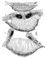
Caption: Fig. 1 Pureke zelandicum. A-C, ascomata in vertical section at different stages of maturity.
(A-C, PDD 49245). | 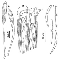
Caption: Fig.2 Pureke zelandicum. A, asci. B, apices of asci and paraphyses. C, released ascospores.
(A-C, PDD 54780). | 
Caption: Fig.7 Macroscopic appearance of ascomata (xl5). Pureke zelandicum (PDD45591). | 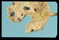
Caption: on Nothofagus menziesii
Owner: Herb. PDD | |
Article: Johnston, P.R. (1991). Rhytismataceae in New Zealand. 4. Pureke zelandicum gen. and sp. nov. plus additional species in Hypoderma, Lophodermium, and Propolis. New Zealand Journal of Botany 29: 395-404 (http://www.rsnz.org/publish/abstracts.php).
Description: Ascomata and structures resembling conidiomata of Rhytismataceae develop in slightly paler
areas of fallen leaves, these areas sometimes surrounded by incomplete, narrow, black zone
lines. Ascomata 0.7-2.0 x 0.4-0-5 mm, broad-elliptic in outline with rounded ends, walls pale
to dark grey, in unopened ascomata with a broad paler zone along the future line of opening,
the outside edge of the ascomata marked by a narrow, black line. Ascomata open by a single
longitudinal slit, the edge of the opening is lined with a narrow, yellowish to dark grey zone.
Ascomata initially subepidermal, but often the epidermal cells becoming filled with fungal
cells as the ascomata mature. In vertical section, at a stage when paraphyses are first
differentiating, the upper wall is 20-30 µm thick, comprising 4-6 rows of hyaline to pale
brown, thin-walled, angular cells. At the same stage the lower wall is 5-15 µm thick,
comprising 1-3 rows of brown to dark brown, thick-walled, angular cells. As the ascomata
mature the upper wall becomes darker and thicker, while the lower wall remains more or less
unchanged. Prior to opening, the upper wall of the ascomata is up to 60 µm thick, slightly
thinner toward the edges, and comprises mostly dark brown, thick-walled, angular cells, but
with a line of pale, thin-walled cells extending through the wall along the future line of
opening. A patch of very dark tissue, in which the cellular structure is obscured, develops in
the inner half of the wall adjacent to the paler cells marking the future line of opening. In
opened ascomata the upper wall is 60-80 µm thick, comprising mostly dark brown, thick-walled, angular cells, but with a patch of very dark tissue in the inner half of the wall near the
ascomatal opening. The exposed face of the broken upper wall is lined with a persistent
palisade-like layer of thin-walled, pale, cylindric cells.
Paraphyses 1.5-2 µm diam., increasing in width suddenly to 6-10 µm at the knob-like apex,
extending 10-15 µm beyond asci. Asci 140-185 x 11-14 µm, clavate-stipitate, tapering to
small, rounded apex, wall undifferentiated at apex, 8-spored, spores confined to upper half of
ascus, development of asci sequential. Ascospores 35-60 x 2.5-3.5 µm, apex rounded,
tapering to base, bifusiform, with abrupt constriction to 1-1.5 µm diam. near centre, 0-1
septate, surrounded by a gelatinous sheath.
Conidiomata-like structures 0.2 mm diam., round in outline, pale brown with darker line
around outside edge, subepidermal. In vertical section lenticular, upper wall lacking, lower
wall 5-10 µm thick, of 1-3 rows of dark brown, thick-walled, angular cells. Conidiogenous
cells and conidia not seen.
Habitat: Fallen leaves of Dacrycarpus dacrydioides, Knightia excelsa, and Nothofagus
menziesii.
Notes: ETYMOLOGY: refers to geographic distribution of this species.
NOTES: The distinctive shape of the ascospores and of the paraphysis tips makes this an easy
species to identify microscopically. It is the only species of Rhytismataceae known from
Dacrycarpus, but it could be confused macroscopically with others on Nothofagus menziesii
(e.g., Lophodermium medium Johnston) and Knightia excelsa (e.g., Lophodermium
brunneolum Johnston).
The collection on Dacrycarpus had longer ascospores (45-60 µm) than all the other
collections (35-45 µm). However, as this was the only morphological difference between the
collections it is considered to represent variation within a single species.
The occurrence of one species on both conifer and angiosperm hosts is unusual in the
Rhytismataceae. However, within New Zealand this kind of host distribution is also seen with
the species Lophodermium agathidis and L. mahuianun (Johnston 1989a).
|