|
 Bivallum zelandicum Bivallum zelandicum
SynonymsBivallum rimu
BiostatusPresent in region - Indigenous. Endemic
Images (click to enlarge)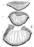
Caption: Fig. 1. Bivallum zelandicum (PDD 49272). A-C, ascomata in vertical section at different
stages of maturity. | 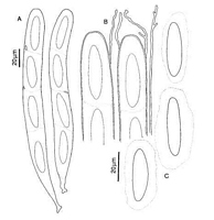
Caption: Fig. 2. Bivallum zelandicum (PDD 49272). A, asci. B, apex of asci and paraphyses. C,
released ascospores. | 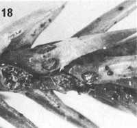
Caption: Fig. 18. Macroscopic appearance of ascomata (scale = 1 mm). Bivallum zelandicum (PDD 56290). | 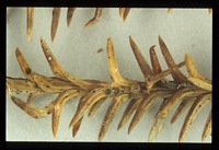
Owner: Herb. PDD | |
Article: Johnston, P.R. (1991). Bivallum gen. nov. (Rhytismataceae) on southern hemisphere Cupressaceae and Podocarpaceae. Australian Systematic Botany 4(2): 355-374.
Description: Ascomata, and in some collections structures which have the appearance of rhytismataceous
conidiomata, develop in pale areas of dead leaves. Not associated with zone lines.
Ascomata in surface view 0.4-0.6 x 0.3-0.4 mm, oblong-elliptic to more or less ovate in
outline. In unopened ascomata, walls pale translucent yellowish grey with dark grey line
marking edge of ascomata. Immediately prior to the ascomata opening a very pale line often
forms along the future line of opening. In opened ascomata the wall is grey to pale grey with
a narrow, dark line marking the outside edge of the ascomata. The single, longitudinal
opening split is lined with a well differentiated, broad, white to yellowish zone.
Ascomata subcuticular. In vertical section at an early stage of ascomatal development, when
paraphyses have started to form but asci are not yet evident (Fig. 1A), the lower wall consists
of 3-4 rows of angular cells, the outermost 1-2 rows thick-walled and dark brown, the inner
rows hyaline and thin-walled. At this stage the upper wall is up to 30 µm wide, mostly of
thin-walled, hyaline, angular cells, although a few cells toward the outside of the wall are
becoming slightly darkened. The inner edge of the wall is lined with short-cylindrical cells
more or less free at their tips. In ascomata where asci are starting to develop (Fig. 1B), the
upper wall is up to 60 µm thick near the centre of the ascomata but becomes suddenly
narrower toward the edges. The inner part of the wide part of the wall is a 10-15 µm wide
layer of narrow-cylindrical, irregularly branching and somewhat tangled hyphae. The outer
part of this central part of the wall is of pale brown, thin-walled, angular cells. The narrower
parts of the wall are of darker brown, slightly thick-walled, angular cells. The opening split
forms through the central, paler part of the wall. The split starts from the outside of the wall,
and as the wall breaks a palisade-like layer of hyaline, cylindrical, unbranched cells develops
across the exposed face of the breaking wall. The wall often starts to break open, and the
layer of cylindrical cells begins to form along the break, prior to the covering host cuticle
breaking. In opened ascomata (Fig. 1 C), the upper wall is up to 80 µm thick near the
opening and there consists of pale brown, thin-walled cells; the rest of the wall is made of
brown, slightly thick-walled, angular cells. The layer of tangled, hyaline cells which was
present toward the inside of the upper wall in unopened ascomata is lost after the ascomata
open. The exposed face of the broken upper wall is lined with a 20-30 µm wide, persistent,
palisade-like layer of hyaline, cylindrical cells. The lower wall now consists of 3-4 layers of
dark brown, thick-walled cells.
Paraphyses 1-2 µm diam., more or less undifferentiated at the apex, extending 5-10 µm
beyond asci. Asci 180-240 x 17-22 µm, saccate with broadly rounded apex, wall
undifferentiated at apex, 4-spored at maturity, with 4 spores aborting. Ascospores 37-44 x 7-11 µm, oblong-elliptic with broadly rounded ends, often slightly constricted near centre,
nonseptate, surrounded by a 5-8 µm wide gelatinous sheath.
Structures resembling conidiomata of Rhytismataceae appear to be associated with ascomata
in some collections. Conidiomata round in outline, 0-2 mm diam., immersed. Conidiogenous
cells and conidia not seen.
Habitat: dead leaves of Dacrydium cupressinum (Podocarpaceae).
Distribution: Known distribution: New Zealand: Northland, Coromandel, Gisborne, Taupo, Buller, Stewart
Is.
Notes: Etymology: refers to the geographic distribution of this species.
Macroscopically and in vertical section B. zelandicum is very similar to the Tasmanian
B. microstrobi.
Coccomyces cupressinum Johnston also occurs on D. cupressinum in New Zealand. C.
cupressinum can be distinguished by its cylindrical asci and filiform ascospores, and ascomata
which usually open by several radiate splits and which are darkened along the edge of the
opening split (Johnston 1986).
Article: Gadgil, P.D. (in association with Dick, M.A.; Hood, I.A.; Pennycook, S.R.) (2005). Fungi on trees and shrubs in New Zealand. Fungi of New Zealand. Ngā Harore o Aotearoa 4: xi + 437 p. Hong Kong: Fungal Diversity Press.
Description: Type: Foliicolous Fungi; Description: Ascomata hysterothecial, scattered, subcuticular, oblong-elliptic to almost oval, pale grey to grey with a narrow dark line marking the outside edge, 0.4–0.6 mm long, opening by a longitudinal slit lined with a broad white to yellowish zone; on both sides of leaves. Asci saccate, 4-spored, 180–240 × 17–22 μm. Ascospores oblong-elliptic, 0-septate, 37–44 × 7–11 μm, surrounded by a 5–8 μm wide gelatinous sheath, hyaline.
Distribution: Distribution: Northland, Coromandel, Taupo, Gisborne, Buller, Stewart Island.; 1st Record: Johnston (1991).
|