|
 Postia venata Postia venata
SynonymsTyromyces venatus
BiostatusPresent in region - Indigenous. Non endemic
Images (click to enlarge)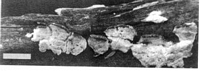
Caption: Fig. 3 Postia venata (MR 10066). Habit. Bar= 1 cm. | 
Caption: Fig. 4 Postia venata (MR 10066), Polyporus setiger (PDD 39360), and Postia atrostrigosa
(PDD 6012). Microscopic characters of the fruit bodies. A-C, P. venata: A, generative hyphae; B,
hymenium; C, spores. D, P. s | 
Caption: Fig. 5 Postia venata (BAFC/cc 575). Aspect of the mat after 6 weeks of growth. | 
Caption: Fig. 6 Postia venata (BAFC/cc 575). Microscopic cultural characters: A, generative hyphae
of the advancing mycelium; B, sclerified, irregularly thickened walled generative hyphae; C,
hyphae with digitiform, sclerified branches. | |
Article: Rajchenberg, M. (1995). Notes on New Zealand polypores (Basidiomycetes) 2. Cultural and morphological studies of selected species. New Zealand Journal of Botany 33(1): 99-109 (http://www.rsnz.org/publish/abstracts.php).
Description: Fruit body annual, effused-reflexed, first forming small circular patches that coalesce, each
patch giving rise to 1-several reflexed portions or pilei that may fuse laterally, remain
separated, or become imbricate. Effused portion of variable size, up to 7 x 6 x 0.4 cm.
Reflexed portion flabelliform or spathulate, first totally white, glabrous or velutinate,
developing more or less abundant strands of dark vinaceous or blackish hairs (Fig. 3). These
strands have a radial disposition, first being aerial and erect,
later totally procumbent on the pileus. The number of strands may increase gradually or
dramatically towards the margin. When mature these strands totally cover the pileus,
resembling veins, with some strands overlapping some of the others and making the pileus
surface black and glabrous. Hymenial surface white and remaining so upon drying. Pores
angular, 4-6 per mm, with entire but irregular and dentate dissepiments. Margin sterile,
incurved. Context white, up to 1.5 mm thick. Tubes white, up to 2.5 mm long, friable upon
drying. Hyphal system monomitic. Generative hyphae clamped, 3-7 µm diam., hyaline, thin-walled
in the growing margins but soon regularly or irregularly thick-walled, swelling in 5%
KOH, acyanophilous, IKI-, giving a positive metachromatic reaction in cresyl-blue. Dark
hairs or veins on the pileal surface formed by dark fuscous hyphae. Tube trama hyphae are up
to 5 µm diam. Basidia cylindric or slightly claviform, with a slight constriction, 12-17 x 3-4
µm. Spores cylindric, some slightly curved, 4-5 x 0.8-1 µm, with thin, hyaline walls, IKI-,
acyanophilous. Cystidia absent (Fig. 4).
Associated wood-rot is chestnut coloured.
CULTURAL STUDIES: Performed with BAFC/cc 575, from specimen M. Rajchenberg
10066 (above).
MACROSCOPIC CHARACTERS (Fig. 5): Growth very slow, up to 4 cm rad. in 6 weeks.
Mat creamy white but poorly developed. Margin regular, appressed, and subfelty. Mat
subfelty, farinose throughout, with slightly marked radial stripes, tightly felty around the
inoculum. Odour: none.
OXIDASE REACTIONS: tannic acid: -, 0 mm; gallic acid: -, trace; tyrosinase: +, 0 mm
MICROSCOPIC CHARACTERS (Fig. 6): Advancing generative hyphae clamped, 4-5 µm
diam., thin-walled, infrequently branched. Older generative hyphae much branched, 1.5-5 µm
diam., with short, digitiform branches that become sclerified; and with irregular wall
thickenings in the wider hyphae. These types of hyphae increase in number towards the
inoculum.
CODE: 1.3c.9.32.36.38.47.54.
Notes: REMARKS: Postia venata was first described from a single collection from southern
Argentina, growing on a fallen trunk of Nothofagus dombeyi (Rajchenberg 1983). Since then
it has been found frequently in the Nothofagus forests of Argentina (Rajchenberg 1993). The
present records from New Zealand expand its distribution (Rajchenberg 1989).
Macroscopically, P. venata may be confused with Tyromyces atrostrigosus (Cooke) G.H.
Cunningham (1965), which also possesses more or less conspicuous tufts of hyphae.
Nevertheless, the latter species differs macroscopically by its strictly flabelliform fruit bodies
and by its context and tubes becoming blue upon drying and handling. Microscopically T.
atrostrigosus differs in its allantoid basidiospores that are wider than 1 µm (Fig. 4E), and by
becoming blue in Melzer's reagent. This species belongs in Postia Fr. because of its
monomitic hyphal system, hyaline spores, clamped generative hyphae that become
metachromatic in cresyl-blue, and the association with a brown wood-rot (David 1980; Rilich
1982).
The following new combination is proposed:
Postia atrostrigosa (Cke) comb. nov. Basionym: Polyporus atrostrigosus Cke,
Grevillea 19: 2. 1890.
Postia atrostrigosa is part of a complex of species surrounding Postia caesia
(Schrad.: Fr.) Karst., given the blue colouration of the fruit bodies upon bruising or
drying and the amyloid, allantoid basidiospores.
Postia venata may also be confused with Tyromyces setiger (Cooke) G.H. Cunn.
(Cunningham 1965; Hood 1992), which possesses allantoid spores and a strigose
pileus. Nevertheless, T. setiger differs in the following characters: fruit bodies
strictly pileate, not effused-reflexed; pilear surface white and uniformly strigose,
with hyaline tufts of hyphae; context up to 30 mm thick and tubes up to 10 mm long;
spores allantoid, 4-5 x 1-1.5 µm (Fig. 4D). I have confirmed these observations in
two collections of T. setiger: New Zealand, Hawke's Bay, Dannevirke, W. Colenso
b517, PDD 39360 (part of type); Auckland, Campbells Bay, on Pittosporum
tenuifolium, J. Dingley, Jul. 1953, PDD 13427. Cunningham (1965) stated that T.
setiger is associated with a white wood-rot. This needs confirmation, as its
morphological features support the inclusion of this species in Postia, a brown wood-rotting genus.
|