|
 Postia globicystidia Postia globicystidia
SynonymsPoria adiposa
Poria albolutescens
BiostatusPresent in region - Indigenous. Endemic
Images (click to enlarge)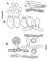
Caption: Fig. 1 Postia globicystidia: A, basidiospores; B, basidia; C, hyphae of context; D, cystidial
elements on context hyphae (PDD 66386 - holotype). A-B, bar = 1C µm; C-D, bars = 10
µm. | 
Caption: Fig. 2 Postia globicystidia: A, habit, showing pore surface (upper left and lower), and
white rhizomorphs on underside of bark (upper right); B, cystidial elements (arrows) on
generative hyphae; C, crystal-encrusted hyphae of rhizomorph (PDD 66 |
Article: Buchanan, P.K.; Ryvarden, L. (1998). New Zealand polypore fungi (Aphyllophorales): three new species and a new record. New Zealand Journal of Botany 36(2): 219-231 (http://www.rsnz.org/publish/abstracts.php).
Description: BASIDIOCARPS annual, resupinate, to 8 x 10 cm, to 2.5 mm thick; pore surface cream to
pale yellow-brown (73.p.OY); margin white, thinning out and in places extended into white
branched rhizomorphs; rhizomorphs also developing from context and then ramifying
under bark of host, to 0.5 mm diam.; pores angular, sometimes with velutinate mouths,
mostly 2-5 per mm but in places much wider, to 2 mm across especially when developing
on near-vertical surfaces; tubes to 2 mm long, concolorous with pores; context white,
narrow, of loose composition. HYPHAL SYSTEM monomitic; generative hyphae hyaline,
clamped, IKI-, non-metachromatic in cresyl blue, non-cyanophilous, 2.5-4 µm diam., rarely
with portions inflated to 8 µm, with wall thin or thickened to 0.5 µm, with encrusting
crystalline material, en crustation sometimes particularly dense on hyphae of the
rhizomorphs and in some specimens present on all hyphae except those of the
subhymenium and hymenium. CYSTIDIAL ELEMENTS arising directly at right angles
from generative hyphae and subtended at base by a clamp, consisting of a thin-walled
clavate structure with apex inflated, resembling immature basidia, e.g., 14-21 x 6.5-10.5
µm, supporting a refractive, globose to subglobose head of dense resinous or wax-like
material 9-15 µm diam. apparently enclosed within a membrane, in most specimens
abundant on hyphae in context and also present, though smaller and less well developed, on
hyphae in trama and in rhizomorphs; supporting cell sometimes lacking a resinous head,
possibly representing an immature stage. BASIDIA broadly clavate to ellipsoid, 4-sterigmate,
basally clamped, 12.5-18 x 6-8 µm. BASIDIOSPORES subglobose, hyaline,
smooth- and thin-walled, IKI-, with prominent apiculus, 4-5(-6) x 3.5-4.3 µm. WOOD
ROT brown.
Habitat: SUBSTRATA: Known from dead wood of Agathis australis, Dacrydium cupressinum,
and Weinmannia racemosa.
Distribution: Northland, Auckland, Stewart Island.
Notes: ETYMOLOGY: globicystidia, referring to the globose to subglobose head of cystidial elements arising from generative hyphae.
NOTES: Cunningham (1965) included specimens of this species among material which he identified as Poria albolutescens (= Anomoporia albolutescens (Romell) Pouzar). Postia globicystidia shares several features with A. albolutescens including small ellipsoid spores, crystal-encrusted hyphae, and a brown rot (Ryvarden & Gilbertson 1993), but differs in having inamyloid spores, white rather than distinctly yellow rhizomorphs, and resinous cystidial elements. Cunningham's concept of A. albolutescens is based on discordant elements. Among his cited material we have also identified the white rot species Ceriporiopsis myceliosa (Peck) Ryvarden & Gilb. and a species of Antrodia P.Karst. or Diplomitoporus.
The cystidial elements (Fig. 1D, 2B) are a conspicuous feature of recent collections (e.g., PDD 53835, 53440, 66386) but are rare in older material (e.g., PDD 7755, 10983, 13456). They are most readily seen in the context, but also may occur in the trama and on hyphae of rhizomorphs. The nature of the apical resinous material is unknown, but it appears to be wax-like in KOH (and may crack when mounted under pressure), resinous in lactophenol, and dissolves or dissipates in Melzers' reagent, leaving behind an empty sphere bounded by an apparent membrane. The overall structure superficially resembles a stephanocyst, though lacking the characteristic frill of teeth at the apex of the supporting cell. In form it is similar to resinous cystidial elements described for species of Hvphoderma Wallr. (Eriksson & Ryvarden 1975, pp. 502, 510). Heavy encrustation of hyphae (Fig. 2C) in collections such as PDD 13456 may obscure these elements. Crystalline encrustation on hyphae is insoluble in KOH and Melzers' reagent.
|