|
 Postia brunnea Postia brunnea
SynonymsGrifola campyla
Grifola rosularis
Polyporus rosularis
Grifola sp. [fig. 84]
BiostatusPresent in region - Indigenous. Non endemic
Images (click to enlarge)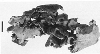
Caption: Fig. 1. Postia brunnea. Dried fruit-body (bar = 1 cm). PDD 20252. | 
Caption: Fig. 2. Postia brunnea. (A) Basidiospores; (B) basidia; (C) chlarnydospores; (D) hyphae
from context. (A-C) material mounted in 3% KOH; (D) material mounted in lactophenol. (A,
B, D) PDD 20252; (C) MR 10094. |
Article: Rajchenberg, M.; Buchanan, P.K. (1996). Two newly described polypores from Australasia and southern South America. Australian Systematic Botany 9(6): 877-885.
Description: Fruit-body annual, lignicolous, compound, with several imbricate pilei or numerous pilei
arising from a common base. Pilei flabelliform, up to 6 x 5 x 0.8 cm, with an attenuated,
narrow base either strictly lateral or elevated, fruit-body somewhat pendent. Margin regular or
irregular, slightly undulated, lacking a sterile portion both at the growing margin and in
lateral parts. Pileus surface continuous laterally with the tubes, pubescent or glabrous,
delicately granulose or roughly pulverulent and, in sectors, with bundles of longitudinally
tightly arranged hyphae; surface longitudinally crenulated, azonate, brown, deep brown to
dark umbrinous chestnut, contrasting with the white hymenial surface and the white to pale
yellow lateral edges of the tubes.
Context white, up to 3 mm thick. Tubes white, up to 5 mm long. Pores regular, round, 3.5-4.5
per mm, with smooth or slightly fimbriate mouths.
Hyphal system monomitic. Generative hyphae with clamps, 5-8 µm diam. in the context, 3-5
µm diam. in the dissepiments, with hyaline walls to 1.5 µm thick, strongly metachromatic in
cresyl-blue, IKI-, swelling and distorting in KOH to leave an irregular lumen that stains
weakly with phloxine. Gloeopleurous hyphae 5-8 µm diam. with contents chestnut or strongly
staining with phloxine.
Basidia claviform, 9-17 x 4-6 µm, 2-4 sterigmate. Spores ellipsoid, 4.0-5.5 x 2.5-3.5 µm,
with an oil-like droplet in the cytoplasm and with slightly thickened, hyaline walls, IKI-;
spores abundant in all collections examined. Chlamydospores globose or ellipsoid, 4-11 µm
diam., with thickened walls, present in the context and the dissepiments of some specimens
(PDD 13375, PDD 6015, PDD 65317; vide infra).
Wood-rot brown.
Notes: Etymology: The specific epithet brunnea means brown, and refers to the coloration of the
pileus surface.
Remarks: Cunningham (1965) refered material of P. brunnea to two species that he
described in the genus Grifola, G. campyla (Berk.) G.Cunn. and G. rosularis (G.Cunn.)
G.Cunn. His descriptions of both of these species were based in part on specimens of
P. brunnea. By comparing an isotype specimen (PDD 28027) of G. campyla
(basionym: Polyporus campylus Berk. (Berkeley 1860)) and the holotype (PDD 3914)
of G. rosularis (basionym: Polyporus rosularis G.Cunn. (Cuuningham 1948)), it was
concluded that these species are conspecific. The recombinatiom Ryvardenia campyla
(Berk.) Rajchenb. (Rajchenberg 1994) is accepted.
Ryvardenia campyla differs from P. brunnea in its dimidiate to flabelliform pilei, the
white to very pale brown coloration of the pileus surface, and the pinkish-beige
marginal area of way texture when fresh. Also, the upper pileus surface has a poroid
sterile structure towards the base. Generative hyphae in R. campyla are thin-walled and
sclerified and then refractive in KOH; they do not swell in KOH, and are not
metachromatic in cresyl-blue. The dissepiments are dimitic with skeletal hyphae.
Spores are hyaline and slightly larger than those of P. brunnea, 4.6-6.5 x 3.7-4.5 µm.
Postia brunnea appears to be closely related to P. pelliculosa (Berk.) Rajchenb.
(Rajchenberg 1988). The species have spores of similar shape and with somewhat
thickened walls. The latter character is unusual in Postia Fr., where most species have
thin-walled spores (Julich 1982). Postia pelliculosa differs from P. brunnea in its
solitary, dimidiate and thickly strigose pelei, sterile margin, pinkish-ochraceous context
and slightly larger spores, 5-7.5 x 3-4 µm. For a description of P. pelliculosa see
Cunnginham (1965) (under Tyromyes), Buchanan & Hood (1992), Hood (1992), Marks
et al. (1982) (under Tyromyces) and Wright and Deschamps (1972) (as Spongipellis
chubutensis Wright & J.R.Deschamps).
Hood (1992) included a brief description and line drawings (figs 84a-e) of P. brunnea,
as Grifola sp. Hood's fig. 84a is a habit drawing of NZFRI 3251, somewhat atypical
because the pelei are immature with pores poorly developed. The fungus photographed
in fig 84 (plate 5) appears to be a different species.
|