|
 Poculum subcinnabarinum Poculum subcinnabarinum
SynonymsHelotium subcinnabarinum
Ciboriella subcinnabarina
BiostatusPresent in region - Indigenous. Endemic
Images (click to enlarge)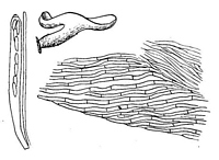
Caption: FIG. 25. Helotium subcinnabarinum. Habit sketch x 12, details x 660. | 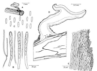
Caption: Figure 47. Poculum subcinnabarinum, holotype. A. Habit sketch. B. Asci and paraphyses.
C. Ascospores. D. Vertical section, showing stromatic development. E. Ectal excipulum on
stipe. | 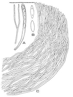
Caption: FIG. 7. Poculum subcinnabarinum, PDD 19052, ex K, camera lucida drawings of median
longitudinal sections of apothecia. A. Paraphysis (right) and ascus (left) with ascospores
biseriate above and uniseriate below, x 1,000. B. Ascospores, x2,000 | 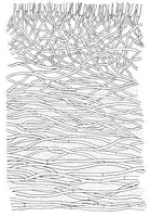
Caption: FIG. 8. Poculum subcinnabarinum, PDD 19052 ex K, camera lucidia drawing, x 1,000.
Median longitudinal section of apothecium at midpoint between margin and stipe. Stippling
represents gelatinous matrix. | 
Caption: FIG. 9. Poculum subcinnabarinum, PDD 19052 ex K, camera lucida drawing, x 1,000.
Median longitudinal section of apothecium at midpoint of stipe. Stippling represents
gelatinous matrix. |
Article: Spooner, B.M. (1987). Helotiales of Australasia: Geoglossaceae, Orbiliaceae, Sclerotiniaceae, Hyaloscyphaceae. Bibliotheca Mycologica 116: 711 p.
Description: STROMA substratal, evident as a superficial blackening of the substrate and, in transverse
section, as two concentric blackened zones surrounding the core of the midrib and petiole.
APOTHECIA hypophyllous, usually arising from petioles or main veins, scattered, superficial,
stipitate. DISC 1.0-2.5 mm diam., planoconcave, or centrally convex and depressed towards
the margin. reddish-orange, smooth. RECEPTACLE shallow cupulate to discoid, paler than
the disc, orange-yellow, smooth. STIPE central concolorous, equalling disc diam., cylindrical
or tapered, with a few longitudinal furrows, smooth above, whitish-tomentose at the base,
with a pad of whitish or pale buff hyphae radiating onto the substrate. ASCI 8-spored, mostly
70-80 x 6-7 µm, cylindric-clavate. tapered below to a rather stout base 3-4 µm diam., apex
rounded or conical, the small pore weakly outlined blue in Melzer's reagent. ASCOSPORES
hyaline, ellipso-cylindric or cylindric-clavate, ends narrowed but rounded, smooth, non-septate, often containing 2 small guttules. obliquely uniseriate or partially biseriate, 8-11 x 2.5-3.0, mean 9.7 (SD 0.7) x mean 2.6 (SD 0.1) µm. PARAPHYSES hyaline, filiform, obtuse,
sparsely septate. often branched near the base, equal to the asci, c. 1.5 µm diam., not
expanding apically. SUBHYMENIUM a poorly differentiated layer 30-40 µm deep, of
subhyaline hyphae 1-5-2.5 µm diam. MEDULLARY EXCIPULUM composed of hyaline or
pale pigmented hyphae 2-3 µm diam., with thin or slightly thickened walls, forming a textura
porrecta in the stipe and in an irregular layer adjacent to the ectal tissue in the receptacle, and
becoming loosely interwoven in the centre of the receptacle, the whole tissue staining blue in
Melzer's reagent, most distinctly in the stipe and lower receptacle. ECTAL EXCIPULUM 30-50 µm thick, composed of parallel, septate hyphae arranged at a low angle to the surface, their
walls thickened and gelatinized. On the stipe, these form thick-walled prismatic cells 20-35 x
5-6 µm, with the terminal 1 or 2 cells thinner-walled and with conspicuous yellow, granular
contents, these appressed to the surface, or sometimes protruding. At the stipe base the ectal
hyphae are more interwoven, forming at the surface interwoven, hyaline hyphae 2-3 µm diam.,
with thin or slightly thickened walls, running out onto the substrate. In the receptacle, the
ectal hyphae are narrower, 4.0-4.5 µm diam., 2-3 µm diam. near the margin, with a few thin
septa, similarly terminating in appressed, sometimes free, thinner-walled elements with
yellowish granular pigment.
Habitat: Typically on leaves of Elaeocarpus dentatus; also on
indet. leaf and wood, and on wood of Nothofagus fusca teste Dumont (1975).
Distribution: New Zealand
Notes: The above description is compiled from the holotype, the only collection of this taxon
preserved in K. Apothecia arise mainly from the petiole and main veins. Stromatic
development is evident as a blackened surface to the substrate, but is also clearly visible in
transverse sections as two concentric black lines surrounding the core of the petiole.
Longitudinal thin sections also reveal narrow, zig-zag black lines through the centre of the
host tissue, and it is clear that the species should be referred to the Sclerotiniaceae. The ectal
excipulum on the receptacle is composed of Interwoven hyphae immersed in a gel, visible as
an unstained refractive layer when mounted in cotton blue, and the species is here considered
to have been correctly placed in Poculum by Dumont (1975). It should be noted that the
medullary excipulum stains distinctly blue throughout in Melzer's reagent, though more deeply
so in the stipe.
This species was one of three referred by Dennis (1964) to Ciboriella, employed for
foliicolous species with reddish tints, lacking stromatic tissue. However, stromatic
development has since been demonstrated in all three species and the genus, as shown by
Dumont (1975), is a synonym of Lanzia. Lanzia griseliniae is similar in appearance to the
present species but lacks a gelatinized excipulum.
Article: Dumont, K.P. (1975). Sclerotiniaceae X. Ciboriella, a taxonomic synonym of Lanzia. Mycologia 67(3): 569-585.
Description: STROMA. Substratal, poorly developed, difficult to detect on host; evident at base of stipe
of apothecium as rind cells; in section epidermoid to irregular in face view, the walls
differentially pigmented. Unknown in culture. MACROCONIDIAL STATE. Unknown.
SI'ERMATIA. Unknown. APOTHECIAL MORPHOLOGY. Apothecia solitary to
gregarious, 1-2.5 mm in diam, to 2 mm high, when fresh flat, English red to Dragon's blood
red, drying cupulate, darker deep orange concolorous with the receptacle and stipe;
rehydrating slightly convex to slightly cupulate, lighter, concolorous, toward base of stipe,
appearing woolly. APOTHECIAL ANATOMY. Asci 8-spored, 67-75(-85) x 5-6(-7) µm,
produced from small croziers, cylindrical to slightly clavate, tapering slightly below to the
base and there expanded and forming a small foot or not; wall narrow, to 1 µm, becoming
thicker at the rounded to slightly truncate apex; pore J+, evident as two tiny blue dots in
Melzer's reagent. Ascospores (7-)8-10 x 2-3 µm, biseriate above and uniseriate below or
tiniseriate throughout, hyaline, fusoid to clavate to ellipsoid, in outline generally slightly
broader at anterior end than posterior, equilateral to slightly inequilateral, with guttules
generally poorly defined, rarely with two, tiny, obvious, internal, polar guttules, more
commonly with two internal polar guttulate areas; walls thin or Appearing thickened in
phloxine stain. Paraphyses equal to or slightly exceeding the asci, very abundant, branching
and septate, filiform, not expanded at the apex and there 1.5-2 µm wide, hyaline, smooth, few
staining intensely and appearing granular internally. Subhymenium a poorly defined zone
beneath the asci, with a zone to ca. 60 µm wide toward the middle of the apothecium and
narrower toward the margin, with hyphae narrower and more compact than the medullary
excipulum below; composed of narrow, loosely interwoven, vertically oriented hyphae 2-3
µm wide, with hyaline and slightly thickened walls. Medullary excipulum well developed,
not refractive or only slightly so in certain areas, consisting of loosely interwoven, branched,
septate hyphae 2-3 µm wide, with several coalescing especially toward the ectal excipulum,
and with smooth, hyaline and slightly thickened walls. Ectal excipulum: inner ectal
excipulum poorly defined, but with a slightly refractive zone of narrow and more compact
hyphae between the medullary excipultim and the outer ectal excipulum, the individual
hyphae 2-3 µm wide with walls smooth, hyaline and slightly thickened. Outer ectal
excipulum highly refractive, tapering from ca. 30 µm broad toward the stipe to ca. 45 µm
broad toward the margin; consisting of narrow hyphae 2-4 µm wide, imbedded in a gelatinous
matrix, extending more or less parallel to each other or slightly interwoven and extending
parallel or at low angles to the surface of the apothecium, the individual hyphae septate,
hyaline, branched and lacking roughened or pigmented walls. Outer covering layer poorly
differentiated from the outer ectal excipulum, not detaching from the surface of the
apothecium, composed of small-celled textura prismatica, 1-2 layers of golden hyphae with
hyaline to subhyaline and slightly thickened walls, lacking any modification of the apical
cells, hairs absent. Margin constructed similarly to the flank, with the individual hyphae
narrower, the spaces between individual hyphae smaller and the gelatinous matrix less
obvious. Stipe constructed similarly to the receptacle, hyphae in the
central core becoming slightly to obviously interwoven.
Habitat: On unidentified leaves and leaves of Elaeocarpus dentatus Vahl, and unidentified
wood and wood of Nothofagus fusca Oest.
Notes: When Dennis (1961) described Helotium subcinnabarinum , he indicated that the
structure of the sterile tissue of the apothecium was somewhat intermediate between
Helotium and Phialea and that the ectal excipulum was composed of interwoven hyphae with
gelatinized walls and extending at a low angle to the surface of the apothecium. My
interpretation is that the hyphae are imbedded in a gelatinous matrix and that structurally it is
more similar to Calycella and Phaeohelotium than either Phialea or Helotium.
I have detected, with some difficulty, rind cells at the base of the apothecium imbedded in the
host. I have also noted the presence of rind cells on the host midvein some distance from the
base of the stipe of the apothecium. Because of the presence of rind cells, characteristic of a
stroma, I refer this species to the Scelerotiniaceae and to the genus Poculum as defined by
Dumont (1972). If it can be demonstrated that this species does not, in fact, produce a stroma
it should be placed in the Leotiaceae, tribe Leotieae.
Article: Dennis, R.W.G. (1961). Some inoperculate Discomycetes from New Zealand. Kew Bulletin 15(2): 293-320.
Notes: The structure is somewhat intermediate between that of an Helotiun and a Phialea, with the
flesh composed of slender interwoven hyphae with slightly gelatinised walls, the hyphae
becoming almost parallel but still undulating at a low angle to the surface in the excipulum.
The red pigment is unaffected by ammonia but completely decolorised by lacto-phenol.
Article: Dennis, R.W.G. (1964). Remarks on the genus Hymenoscyphus S.F. Gray, with observations on sundry species referred by Saccardo and others to the genera Helotium, Pezizella or Phialea. Persoonia 3(1): 29-80.
Description: Spores 8-9 x 2-2.5 µ.
Notes: Ciboriella Seaver, North Amer. Cup Fungi (Inop.) I07. 195I.
Soft-fleshed species with reddish tints, reminiscent of Sclerotiniaceae but with an excipulum
composed of short-celled parallel hyphae and with no sclerotium or stromatic tissue; asci I+,
on dead leaves.
|