|
 Meloderma dracophylli Meloderma dracophylli
BiostatusPresent in region - Indigenous. Endemic
Images (click to enlarge)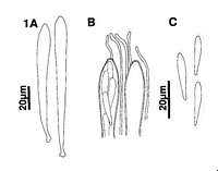
Caption: Fig 1 Meloderma dracophylli. A. Asci. B. Detail of ascus and paraphyses apex. C. Released
ascospores. | 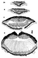
Caption: Fig 2 Meloderma dracophylli (PDD 45303), ascoscarps in vertical section. A. Immature ascocarp prior to development of paraphyses. B, C.
Unopened ascocarps showing internal differentiation of upper wall. D. Upper wall of ascocarp starting to bre | 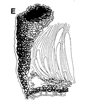
Caption: Fig 2 Meloderma dracophylli (PDD 45303), ascoscarps in vertical section. A. Immature ascocarp prior to development of paraphyses. B, C.
Unopened ascocarps showing internal differentiation of upper wall. D. Upper wall of ascocarp starting to bre | 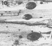
Caption: Fig 10 Macroscopic appearance of ascocarps (x 15). Meloderma dracophylli (PDD 44481). | 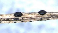
Caption: Ascomata about 0.7 mm long.
Owner: Herb PDD |
Article: Johnston, P.R. (1988). A new species of Meloderma (Rhytismataceae), with notes on Meloderma and related genera. Mycotaxon 33: 423-436.
Description: Ascocarps and pycnidia developing in pale areas of fallen leaves, often surrounded by narrow,
black zone lines. Ascocarps 0.5-0.8 x 0.4-0.5 mm, broad-elliptic to more or less circular in
outline. Immature, unopened ascocarps with uniformly black walls. When mature, walls
oriented at a steep angle to surrounding host tissue, black, with a flattened, pale area around
the single opening slit. Opening not extending to ends of ascocarp, and often with the two
sides of the opening slit pulling apart so exposing the hymenium even when ascocarp dry.
Pycnidia 0.2 mm, circular in outline, brown-walled, pustulate.
Ascocarps subcuticular. In vertical section upper wall up to 100 µm wide near ascocarp
opening, narrower toward outer edge. Upper wall comprising 2 layers, an outer layer of dark
brown, thick-walled, angular cells, and an inner layer of hyaline, thin-walled, downward-projecting, cylindric cells. The cells in part of the inner layer near the ascocarp opening
becoming dark brown and thick-walled as the ascocarp matures. Exposed face of broken
upper wall lined with layer of short-cylindric, hyaline to pale brown cells. Lower wall 10-15
µm wide, of 2-3 layers of brown to dark brown, thick-walled cells.
Paraphyses 1.5-2 µm diam., often circinate near apex, extending 10-15 µm beyond asci.
Asci 95-125 x 9-12.5 µm, clavate, tapering gradually to base, and tapering to small, truncate
apex, 8-spored. Ascospores 19-25 x 3.5-4 mm, narrowly obovoid, tapering toward base, ends
rounded, 0 septate, surrounded by a well-developed gelatinous sheath.
Pycnidia in vertical section subcuticular, upper wall 5 µm wide, comprising very dark tissue
with no obvious cellular structure. Lower wall 10-15 µm wide, comprising dark brown, thick-walled, angular cells lined with conidiogenous layer. Conidiogenous cells 15-22 x 2-3 µm,
tapering to apex, solitary, with sympodial proliferation, often with 2 conidia held at apex.
Conidia 3.5-5 x 1-1.5 µm, cylindric with rounded ends, 0 septate. Amongst conidiogenous
cells are filiform sterile elements up to 45 µm long.
CHARACTERISTICS IN CULTURE: Ascospores germinating on agar plates within 48
hours. On oatmeal agar colonies 4.5 mm diam. after 6 weeks; aerial mycelium low, sparse,
cottony, white. agar surface with pinkish, vinaceous, or yellowish colours toward centre of
colony. Black-walled, globose pycnidia on agar surface, opening by an irregular slit to expose
conidial ooze, conidiogenous cells and conidia as described in vivo.
Habitat: On dead, fallen leaves of Dracophyllum longifolium, D. uniflorum Hook.f, D.
subulatum Hook.f.
Distribution: New Zealand: Taupo, Gisborne, Taranaki, Buller, North Canterbury, Mid
Canterbury.
Notes: ETYMOLOGY: dracophylli; refers to host substrate.
NOTES: M. dracophylli is macroscopically indistinguishable from the Australian species M.
richeae. However, M. richeae has shorter, 4-spored asci, and larger, often 1-septate
ascospores.
|