|
 Lanzia griseliniae Lanzia griseliniae
SynonymsHelotium griseliniae
Ciboriella griseliniae
BiostatusPresent in region - Indigenous. Endemic
Images (click to enlarge)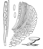
Caption: FIG. 19. Helotium griseliniae. Habit sketch x 7, details x 660. | 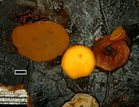
Caption: scale=1mm
Owner: J.A. Cooper | 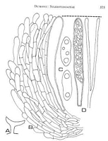
Caption: FIG. 4. Lanzia griseliniae, PDD 19046 ex K, camera lucida drawings of median longitudinal sections
of apothecia. A, whole apothecium, ca. x10. B. Margin, x1000. C. Ascospores, x2,000. D. Ascus (left) wth ascospores uniseriate and paraphyses
(ri | 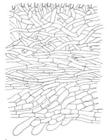
Caption: FIG. 5. Lanzia griseliniae, PDD 19046 ex K, camera lucida drawing, x 1,000. Median
longitudinal section of apothecium at approximately midpoint between margin and stipe. | 
Caption: FIG. 6. Lanzia griseliniae, PDD 19046 ex K, camera lucida drawing, X 1,000. Median
longitudinal section of apothecium at approximately midpoint of stipe. | 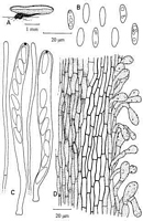
Caption: Figure 66. Lanzia griseliniae, holotype. A. Apothecium. B. Ascospores. C. Asci and
paraphyses. D. Ectal excipulum on stipe. |
Article: Spooner, B.M. (1987). Helotiales of Australasia: Geoglossaceae, Orbiliaceae, Sclerotiniaceae, Hyaloscyphaceae. Bibliotheca Mycologica 116: 711 p.
Description: STROMA substratal, poorly developed, evident as slight blackening of the leaf surface in
places, and weak stromatic lines. APOTHECIA hypophyllous, erumpent, stipitate, solitary.
DISC 1.5-3.0 mm diam., shallow concave or depressed around a convex centre, smooth,
purple-brown when fresh (Dennis, 1961), dull orange-brown to brick red when dry,
rehydrating orange. RECEPTACLE cupulate, reddish brown, slightly darker than the disc,
and darker towards the stipe, minutely scurfy except at the extreme margin. STIPE central,
cylindric, usually shorter than disc diameter, concolorous or dark brown when rehydrated,
curved, distinctly scurfy. ASCI 8-spored, cylindric-clavate, tapered below to a short stalk-like
base, apex broadly rounded, measuring (95-)100-120 x 7.5-9.5 µm, pore up to 2 µm deep,
outlined strongly blue in Melzer's reagent. ASCOSPORES ellipsoid, broadest above centre,
often inequilateral, hyaline, containing 2 large or several smaller guttules, non-septate,
obliquely uniseriate or partially biseriate, 11.5-14.5 x 3.5-4.8, mean 12.8 (SD 0.8) x mean 4.2
(SD 0.3) µm. PARAPHYSES filiform, obtuse, hyaline, rarely septate, unbranched, equal to
the asci, 1.5-2.0 (-2.5) µm diam., slightly enlarged towards the apex to 2-3 µm diam.
SUBHYMENIUM indistinct, c. 20 µm thick, composed of loosely interwoven, vertically
orientated subhyaline hyphae 2.5-4.0 µm diam. MEDULLARY EXCIPULUM composed in
the receptacle of interwoven, hyaline or usually pale yellowish-brown thin-walled hyphae 4-6
µm diam., merging with the medulla of the stipe in which the hyphae are more compact,
vertically arranged, pale brown, 3.5-4.5 µm diam., the walls granularly roughened due to
irregular deposition of pigment. ECTAL EXCIPULUM in stipe and lower receptacle
composed of hyaline prismatic cells, often with slightly thickened but not refractive walls, 13-18(-25) x 4-7 µm, forming a layer 20-35 µm thick, overlying a zone of vertically orientated
brown to dark brown septate hyphae 3-5 µm diam. This zone is up to about 50 µm wide
towards the stipe base, narrowing to 20 µm at the stipe apex, and disappearing in the
receptacle. Ectal layer in receptacle 20-30 µm thick composed of pale brown, thin walled
prismatic cells mostly 12-18 x 5-7 µm, arranged in rows at a low angle to the surface, longer
and narrower at the margin. At the surface throughout, but more conspicuously developed on
the stipe, the ectal cells are pigmented and give rise to free, hair-like extensions, clavate and
often strongly expanded at the tips, constricted at the septa, dark brown at the base of the
stipe, there up to 50 µm long, (5-)10-15 µm diam., with irregularly deposited often granular
pigment. Towards the margin these elements are shorter, paler and less conspicuous, and no
more than free, clavate hyphal tips 5-10 µm diam. These structures do not form a uniform
surface layer on the stipe, but cohere irregularly to give the receptacle a scurfy appearance
under a lens rather than a uniform downy covering
Habitat: On leaves of Griselinia littoralis. New Zealand, known
only from the type locality.
Notes: This species was described and illustrated by Dennis (1961) as Helotium griseliniae.
Subsequently, Dumont (1975) reported the presence of a black line-stroma in the type
material, and transferred the name to Lanzia. The species is known only from the type
collection, which consists of two leaves bearing several well-developed apothecia erumpent
from the underside of the blades. In one leaf, a general blackening of the surface is apparently
caused by a sooty mould and the only clear line-stroma delimits an area at the edge of the
blade remote from the apothecia. In the other, a line stroma runs across the blade above the
petiole though cannot be shown to be positively associated with the apothecia. However, a
slight stromatic development which does seem to be associated with an apothecium is present
near the blade edge. The structure and pigmentation of the apothecia also strongly suggests
that this species belongs in the Sclerotiniaceae. The species is probably best placed in Lanzia.
However, until fresh material is available and can be successfully grown in culture, the
existence and characters of a stroma must remain uncertain.
There are several foliicolous species currently referred either to Lanzia or Rutstroemia
which may be closely allied to L. griseliniae. These include Lanzia longipes (Cooke & Peck)
Dumont & Korf, and L. luteovirescens (Roberge) Dumont & Korf (Dumont, 1975). Both are
petiolicolous; the former lacks the characteristic pigmented surface hyphae of L. griseliniae,
whereas the latter differs particularly in colour and in having broader ascospores. Rutstroemia
pruni-serotinae Whetzel & White occurs on the leaf blade and produces conspicuous black
stromatic lines. While comparable in colour and possessing a striolate surface to the
receptacle, it has much smaller ascospores and is restricted to leaves of Prunus serotina.
Lanzia rubescens (Kanouse) Dumont, on leaves of species of Alnus in North America has a
similar stromatic development and yellow or orange apothecia which dry to a reddish colour.
It differs from L. griseliniae in having much smaller ascospores and only very short hair-like
hyphae projecting from the surface of the receptacle (Dumont, 1975). Lanzia albo-atra may
be more closely related to the present species. It occurs on dead leaves in Brazil. A black line
stroma is present in this species and the apothecia have an outer layer of hyphae terminating in
apically free cells which are brown and frequently roughened, very similar in most respects to
the structure of L. griseliniae (Dumont, 1981). Lanzia albo-atra differs most notably in
colour, having a white hymenium and receptacle which dry black, and a black stipe, and also in
having broader asci and spores. The characters of the foliicolous L. berggrenii and L.
ovispora are discussed elsewhere in the present account.
Article: Dumont, K.P. (1975). Sclerotiniaceae X. Ciboriella, a taxonomic synonym of Lanzia. Mycologia 67(3): 569-585.
Description: STROMA. Substratal, evident as single or double black lines extending irregularly along the
leaf blade especially toward the margin, in section the black lines consisting of a rind of cells
with differentially pigrnented walls, in surface view the individual cells irregular to
epidermoid. Unknown in culture. MACROCONIDIAL STATE. Unknown. SPERMATIA.
Unknown. APOTHECIAL MORPHOLOGY. Apothecia 1-3 mm in diam, to ca. 1.5 mm
high, when fresh purple-brown (Dennis, 1961), flat to slightly convex, drying cupulate,
hymenium brick red, receptacle purple-black to black, concolorous with the stipe which is
eccentric, tapering toward the base; rehydrating flat and lighter when dry. APOTHECIAL
ANATOMY. Asci 8-spored, (85-)90-110(-125) x 8-10 µm, produced from croziers, cylindric
to slightly clavate, tapering slightly below toward the base and there slightly expanded to
form a small foot; wall 1-2 µm thick, 2-3 µm at the rounded apex; pore J+, the walls outlined
deep blue in Melzer's reagent. Ascospores (11-)12-14(-15) x (3-)4-5 µm, biseriate above and
uniseriate below or uniseriate throughout, hyaline, ellipsoid, smooth, in outline inequilateral,
generally slightly broader at anterior end than posterior, rarely slightly indented and
subreniform; with two, large, internal polar guttules, the anterior one frequently larger than
posterior, occasionally breaking into smaller guttules. Paraphyses slightly exceeding the asci,
hyaline, rarely branched and septate, filiform, 2-3 µm wide at the rounded apex.
Subhymenium to ca. 40 µm broad, whole layer vertically oriented and subliyaline to lightly
pigmented, not refractive; consisting of narrow, tightly interwoven hyphae with thick,
smooth, hyaline to lightly pigniented walls. Medullary excipulum well developed, not
refractive, consisting of loosely to tightly interwoven, branched, septate hyphae 4-7 µm wide,
with frequently coarsely roughened and pigmented walls. Ectal excipulum: inner ectal
excipulum well developed, 3 to several layers broad, tapering from ca. 10 µm broad toward
the margin to ca. 25 µm broad toward the stipe; consisting of parallel to slightly interwoven,
tightly to loosely compact hyphae, 2-5 µm wide, the individual cells shortest and narrowest
toward the margin and longest and broadest toward the stipe, with coarsely roughened and
lightly pigmented walls (roughenings and pigmentation more intense and obvious toward the
margin). Outer ectal excipulum not refractive or only slightly so, tapering from ca. 15 µm
wide toward the margin to ca. 30 µm wide toward the stipe; consisting of a well-developed
textura prismatica of 2-4 parallel hyphal layers extending at low to high angles to the surface
of the apothecium, the individual cells frequently becoming disrupted and losing hyphal
orientation and with hyaline to lightly pigmented and smooth, to coarsely roughened walls
with granular deposits between adjoining cells. Outer covering layer present, generally well
developed, but occasionally becoming detached from the outer surface and appearing naked,
0-2 layers of narrow hyphae with lightly to intensely pigmerited and coarsely roughened
walls; individual hyphae becoming modified apically, with the terminal or the last two to
three cells becoming greatly expanded and constricted at their septa; several apically free tips
often detaching together and appearing scale-like. Hairs absent, but the apical cells of the
hyphae of the outer covering layer become modified and appear hairlike. Margin narrow,
constructed similarly to the upper portion of the apothecial flank, the apically free cells less
expanded. Stipe constructed similarly to lower portion of the flank of the apothecium, the
entire surface clothed with the apically free cells of the outer covering layer.
Habitat: Leaves of Griselinia Iittoralis Raoul.
Distribution: Known only from the type collection.
Notes: When Dennis (1961, 1964) treated L. griseliniae, he failed to detect the black line
stroma present on the leaves of the host plant and placed the species in the Helotiaceae. With
the report here of the presence of a stroma, this species is surely a member of the
Sclerotiniaceae, and is placed in the gentis Lanzia following the concept of Dumont (1972).
This species appears to be most closely related to "Rutstroemia longipes" and "R.
luteovirescens." In R. longipes the apothecia emit a reddish brown to purplish dye in 2%
KOH, while in L. griseliniae a faint yellowish dye is given off. Rutstroemia longipes also
lacks the characteristic outer covering layer of L. griselinae. Lanzia griseliniae is most easily
distinguished from R. luteovirescens by characters of the ascospores. In L. griseliniae the
ascospores are flattened on one side and slightly broader at one end, while in R.
luteovirescens they are rarely slightly inequilateral, but not flattened on one side and both
ends are more or less equal.
Notes: There are several leaf-inhabiting, sclerotiniaceous discomycetes with bright yellow to mustard
coloured apothecia. These include Lanzia griseliniae along with several putatively distinct,
undescribed species. All are macroscopically similar when fresh, bright yellow to mustard
yellow, all have an outer excipular layer with the end cells of the outermost elements free,
swollen, and generally packed with bright yellow pigments, but pigments are not released into
KOH (expect sometimes when apothecia are young, and then only in small quantities). The
colour of the apothecia is variable after drying, often darker, although this varies within a
collection; the apothecia of the species on Myrsine chathamica dry consistently orange. Cells
of the outer exipular layer are short and broad, angular to almost globose, with the wall
thickened. The putative taxa can be distinguished primarily by small variations in ascospore
size. Each of the undescribed taxa are known from two or more collections, with some host or
geographic specialisation, although this not absolute. The morphologically defined taxa need
testing with molecular data.
- Lanzia griseliniae (Dennis) Dumont, on Griselinia, not common, known from
4 collections on Griselinia, the type from the central North Island, others from
Banks Peninsula, Stewart Island, and Karamea, as well as one from
Pseudopanax from Taupo. Ascospores about 12-15 x 4-5 µm; asci about 8-11
µm wide.
- Lanzia sp. "aurea" quite common on Griselinia spp., Pseudopanax
crassifolius and Metrosideros umbellata, with one collections on Senecio
reinoldii. Known from the South Island, Stewart Island and the Auckland
Islands. This species differ from L. griseliniae in having smaller ascospores,
about 9-11 x 3-4 µm; asci about 7-8.5 µm wide.
- Lanzia sp. "aurea pohutukawa" on Metrosideros excelsa. These few
collections differ from Lanzia sp. "aurea" having shorter and wider
ascospores, 7-8.5 x 3.5-4.5 µm.
- Lanzia sp. "aurea chathamica", on Myrsine chathamica, Chatham Islands.
When dry the more or less free cells covering the apothecia of these
collections are bright orange rather than bright yellow (when fresh they are
yellow), and the apothecia release small quantities of orange-red pigments into
KOH (the other taxa release either no pigments, or small quatities of yellow
pigment). Some of the free cells on the outer excipular layer are distinctly
fusoid and apiculate; the hyphae of the outer excipular layer are not encrusted.
The ascopores match those of Lanzia sp. "aurea".
Article: Dennis, R.W.G. (1961). Some inoperculate Discomycetes from New Zealand. Kew Bulletin 15(2): 293-320.
Description: Excipulum_composed of parallel, short-celled hyphae, 7-8 µ wide, with thin, soft, red brown
walls, lying at a low angle to the surface.
Article: Dennis, R.W.G. (1964). Remarks on the genus Hymenoscyphus S.F. Gray, with observations on sundry species referred by Saccardo and others to the genera Helotium, Pezizella or Phialea. Persoonia 3(1): 29-80.
Description: Spores I2-I4 x 4-5 µ.
Notes: Ciboriella Seaver, North Amer. Cup Fungi (Inop.) 107. 1951.
Soft-fleshed species with reddish tints, reminiscent of Sclerotiniaceae but with an excipulum
composed of short-celled parallel hyphae and with no sclerotium or stromatic tissue; asci I+,
on dead leaves.
|