|
 Lanzia berggrenii var. metrosideri Lanzia berggrenii var. metrosideri
SynonymsHelotium metrosideri
Hymenoscyphus metrosideri
BiostatusPresent in region - Indigenous. Endemic
Images (click to enlarge)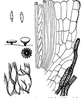
Caption: FIG. 20. Helotium metrosideri. Habit sketches x 10, left, dry; right, soaked up, details x 660,
except for diagram of surface net which is c. x 100. | 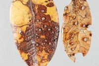
Owner: Herb PDD | 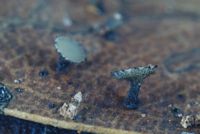
Owner: Herb PDD | 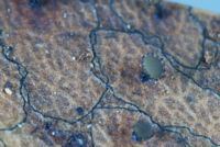
Owner: Herb PDD | 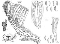
Caption: Figure 68. Lanzia berggrenii var. metrosideri. A-E- Holotype. A. Apothecium. B. Vertical
section, upper receptacle. C. surface hyphae. D. Asci and paraphyses. E. Ascospores. F-G.
Dingley 19379. F. Ascus and paraphyses. G. As |
Article: Spooner, B.M. (1987). Helotiales of Australasia: Geoglossaceae, Orbiliaceae, Sclerotiniaceae, Hyaloscyphaceae. Bibliotheca Mycologica 116: 711 p.
Description: STROMA substratal, visible on both leaf surfaces as dark lines delimiting areas surrounding a
single apothecium, and as dark areas at the base of the apothecia. APOTHECIA scattered or
gregarious, hypophyllous, stipitate, erumpent through the epidermis which is split into 5 or 6
conspicuous erect or reflexed dark tooth-like lobes. DISC 0.7-1.0 mm diam., convex or
plane, smooth, pallid with an olivaceous tinge. RECEPTACLE reflexed, pale, covered by a
conspicuous meshwork of dark brown hyphae extending beyond the margin to form a dentate
fringe to the disc. STIPE central, cylindric, dark brown to blackish, shorter than disc diam.
ASCI 8-spored, cylindric or cylindric-clavate, narrowed below to a very short stalk, apex
broadly conical or truncate, the pore only faintly outlined blue in Melzer's reagent, 80-95(-108) x 8-10 µm. ASCOSPORES hyaline, fusoid, inequilateral, often slightly curved, non-septate, containing a row of 3-5 large guttules, obliquely uniseriate or irregularly biseriate,
13.0-17.0 x 3.5-4.8, mean 15.2 (SD 0.9) x mean 4.2 (SD 0.3) µm. PARAPHYSES filiform,
simple, 1.0-1.2 µm diam., enlarged above to a clavate or sometimes capitate apex, 3.5-5.0 µm
diam., containing pale yellow granules of pigment, exceeding the asci by 5-8 µm.
SUBHYMENIUM not clearly differentiated, composed of loosely woven hyaline hyphae 3-5
µm diam. MEDULLARY EXCIPULUM a narrow layer 30 µm thick at the base of the
receptacle, narrowed to the margin, composed of parallel, subhyaline or pale brown hyphae 3-4 µm diam. In the stipe the hyphae are broader, 6-9 µm diam., vertically arranged, narrower
towards the ectal layer. ECTAL EXCIPULUM composed of large prismatic cells with
thickened walls, 15-20 x 9-14 µm in the receptacle, in rows at a very low angle to the surface.
On the stipe the ectal cells are longer and narrower, 20-30 x 4-7 µm forming a layer not
clearly differentiated from the medullary tissue and frequently with the pigment deposited in
irregular granules. Ectal layer thin, 1-3 cells thick on the receptacle, 20-25 µm thick, giving
rise at the surface to dark brown hyphae 4-7 µm diam. with clavate tips 6-10 µm diam.,
strongly encrusted throughout with dark brown pigment. Surface hyphae more densely
developed at the margin and extending beyond the disc as a dentate marginal fringe. Those on
the receptacle surface irregularly pulled apart as the apothecium expands to form an irregular,
very conspicuous meshwork; those on the stipe narrower, 2.5-3.0 µm diam., the outermost
excipular hyphae also pigmented forming a dark surface layer 5-8 µm thick. At the stipe base,
diverging, dark brown, septate hyphae 3-4 µm diam., bind with the upraised epidermis to form
the conspicuous dark tooth-like lobes surrounding the stipe base.
Habitat: On dead leaves of Metrosideros sp. and M. robusta
(Myrtaceae). Auckland, New Zealand.
Notes: This beautiful and distinctive taxon is known only from two collections from Auckland, New
Zealand. It differs from the type in the more conspicuous meshwork of surface hyphae, which
project beyond the disc as a fringe of marginal teeth, the dark stipe and less flared margin. In
addition, the paraphyses are simple, clavate or capitate at the apex and the asci are slightly
longer and have a less cylindrical form. There are no differences in the ascospore characters.
These differences in the form of the apothecia correlate with a difference in host, which
provides further evidence for regarding these taxa as distinct at varietal level.
Lanzia berggrenii was redescribed and illustrated by Beaton & Weste (1976c) as
Pezizella nothofagi. They have accurately described the asci, paraphyses and excipular
structure, but failed to note the presence of a stroma.
Article: Dennis, R.W.G. (1961). Some inoperculate Discomycetes from New Zealand. Kew Bulletin 15(2): 293-320.
Notes: H. metrosideri is structurally not unlike H. scutula, with flesh of loosely woven hyaline
hyphae, 8-10 µ wide, and excipulum of compact parallel hyphae of similar size, lying at a low
angle to the surface, covered by a thin web of more slender hyphae pulled somewhat apart into
a mesh-work as the apothecium expands on wetting. These surface hyphae, 4-6 µ wide, are
blackened by a granular deposit. There is in this a close resemblance to the structure of H.
spadiceo-atrum (Mont.) Sacc., on Gunnera scabra in Juan Fernandez, but in the latter the
ascospores are only 9-10 x 3 µ and the surface hyphae have a reddish-brown granulation and
yield a purple solution in KOH.
Article: Dennis, R.W.G. (1964). Remarks on the genus Hymenoscyphus S.F. Gray, with observations on sundry species referred by Saccardo and others to the genera Helotium, Pezizella or Phialea. Persoonia 3(1): 29-80.
Description: Spores 14-17 x 4-5 µ.
Notes: SERIES 7. Prasinum. Stipitate species with a large-celled inner excipulum covered by dark
encrusted hyphae or at least with brown walled hyphae covering the stipe
The series shows affinities with Rutstroemia but I think its addition to that genus inadvisable.
|