|
 Hypoderma sticheri Hypoderma sticheri
BiostatusPresent in region - Indigenous. Endemic
Images (click to enlarge)
Caption: Fig. 13 Hypoderma sticheri: A, margin of ascoma in vertical section (PDD 54879). B,
immature ascoma in vertical section (PDD 48328). C, asci (PDD 48328). D, apex of ascus
and paraphyses (PDD 48328). E, released ascospores and ascoconidia (P | 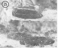
Caption: Fig. 25 Hypoderma sticheri (PDD 43974). Macroscopic appearance of ascomata (x15). |
Article: Johnston, P.R. (1990). Rhytismataceae in New Zealand 3. The genus Hypoderma. New Zealand Journal of Botany 28(2): 159-183 (http://www.rsnz.org/publish/abstracts.php).
Description: Ascomata and conidiomata developing on dead fronds within slightly paler, yellowish areas on
rachises of fronds. Not associated with zone lines. In surface view ascomata 2-3 x 0.6-1.0
mm, oblong to elliptic in outline, pointed to ends. Unopened ascomata with uniformly black
walls. Ascomata opening by single, longitudinal slit, edge of opening lacking any visible
differentiated zone. Conidiomata 0.2-0.3 mm diam., circular in outline, black, pustulate.
Ascomata subcuticular. In vertical section upper wall of unopened ascomata up to 100
µm thick. Outer part of wall comprising dense, black material, in a layer up to 50 µm wide
toward the centre of the ascomata, with the inner part comprising brown to pale brown, thin-walled, angular cells. In a narrow line, along the future line of opening, the paler cells extend
almost to the outside of the wall. A few hyaline, thin-walled, cylindric cells line the inside of
the wall. In opened ascomata the upper wall is up to 110 µm thick near the opening,
becoming thinner toward the base, the outer part and the part of the wall near the ascomatal
opening comprising dense, black tissue with no visible cellular structure; the inner part of pale,
thin-walled, angular cells. The exposed face of the broken wall becomes lined with a layer of
cylindric, hyaline to brown, thin-walled, 15-20 x 3-4 µm cells. Lower wall less than 5 µm
thick, of one layer of dark brown, angular, thick-walled cells.
Paraphyses 1-2 µm diam., undifferentiated at apex, barely extending beyond asci. Asci
120-160 x 17-22 µm, clavate, becoming clavate-stipitate prior to spore release, tapering to
truncate apex, wall slightly thinner across apical part of ascus, 8-spored. Ascospores 24-39 x
3.5-5 µm (average 33.4 x 4.1 µm) bifusiform, 1.5-2.0 µm wide at central constriction, 1-septate, surrounded by gelatinous sheath. Ascospores within the ascus often breaking across
the central septum into two part-spores. When this occurs ascoconidia are produced from the
broken end of each part-spore, and the asci eventually become full of the 4.0-7.5 x 1.0-1.5
µm, cylindric, 0-septate, hyaline ascoconidia.
Conidiomata subcuticular. In vertical section lenticular in shape. Upper wall 5-10 µm
thick, comprising dark brown tissue with noo bvious cellular structure. Lower wall 5-15 µm
thick, of 1-3 rows of dark brown, thick-walled, angular cells. Conidiogenous layer develops
on lower wall. Conidiogenous cells 8-13 x 1.5-2.5 µm, cylindric, tapering near apex, wall
slightly thickened at the single, apical conidiogenous locus, proliferation percurrent. Conidia
3.5-4.5 x 1.0-1.5 µm, oblong-elliptic, 0-septate, hyaline.
Habitat: On midribs of dead fronds of Sticherus cunninghamii (= Gleichenia
cunninghamii).
Distribution: Taupo, Buller, Westland.
Notes: ETYMOLOGY: named after host substrate.
NOTES: This is the only Hypoderma species in New Zealand known from a fern. It is easily
distinguished from other species by its large ascomata, black-walled conidiomata, large asci,
and by its 1-septate, often disarticulating ascospores. The appearance of the upper wall of
unopened ascomata is distinctive with its broad inner layer of pale, thin-walled cells.
|