|
 Hypoderma sigmoideum Hypoderma sigmoideum
BiostatusPresent in region - Indigenous. Endemic
Images (click to enlarge)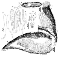
Caption: Fig. 12 Hypoderma sigmoideum: A, margin of ascoma in vertical section (PDD46212). B,
immature ascoma in vertical section (PDD 46212). C, asci (PDD 46212). D, apex of asci and
paraphyses (PDD 46212). E, released ascospores (PDD 46212). F, c | 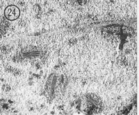
Caption: Fig. 24 Hypoderma sigmoideum (PDD 46212). Macroscopic appearance of ascomata (x15). | 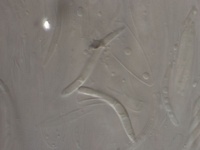
Caption: sigmoid ascospores
Owner: P.R. Johnston | 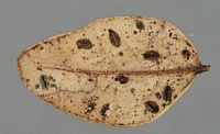
Caption: characteristic red lip cells
Owner: P.R. Johnston | 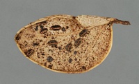
Caption: characteristic red lip cells
Owner: P.R. Johnston |
Article: Johnston, P.R. (1990). Rhytismataceae in New Zealand 3. The genus Hypoderma. New Zealand Journal of Botany 28(2): 159-183 (http://www.rsnz.org/publish/abstracts.php).
Description: Ascomata and conidiomata developing on fallen leaves within pale whitish or yellowish areas
on host leaf This pale area often surrounded by narrow, black zone lines. In surface view
ascomata 1.0-1.5 x 0.6-0.7 µm, broad-elliptic in outline, ends rounded. Unopened ascomata
black-walled. Ascomatal opening lined with narrow, bright orange or red zone, or if lined
with yellow zone then this changes to red when treated with 3% KOH. Conidiomata 0.2-0.3
mm diam., circular or slightly irregular in outline, black walled, pustulate.
Ascomata subcuticular. In vertical section upper wall of unopened ascomata up to 70 µm
thick, narrower toward edges, comprising mostly brown to dark brown, angular cells. Cells
darker and thicker walled along outer edge of the wall, and along most of the inside edge.
Along the future line of opening, toward the inside of the upper wall, is a group of paler,
thinner-walled cells. In opened ascomata the upper wall is up to 70 µm thick near the
ascomatal opening, becoming gradually thinner toward the base of the wall. Upper wall
comprising brown to dark brown, thick-walled, angular, 4-8 µm diam. cells. Exposed face of
the broken upper wall is lined with cylindric, hyaline, thin-walled, 30-45 x 4-5 µm cells.
Lower wall 5-10 µm thick, of 2-3 layers of brown, thick-walled, angular, 4-7 µm diam. cells.
Paraphyses 1-2 µm diam., circinate at apex, extending 5-10 µm beyond asci. Asci 115-145 x 9.5-13.0 µm, clavate-stipitate, tapering to rounded apex, wall undifferentiated at apex,
8-spored, spores confined to upper half of ascus. Ascospores 22-38 x 3-4 µm (average 30.6 x
3.3 µm), sigmoid, tapering to base, 1-septate, surrounded by a narrow gelatinous sheath.
Conidiomata subcuticular. In vertical section upper wall 5 µm thick, comprising dense,
dark brown material with no obvious cellular structure. Lower wall 10 µm thick, of 2-3 rows
of brown, slightly thick-walled, angular to cylindric, 4-6 µm diam. cells. Lower wall is lined
with 2-3 lavers of hyaline, thin-walled, angular, 3-4 µm diam. cells on which the
conidiogenous cells develop. Conidiogenous cells 10-17 x 1.5-2.5 µm, cylindric or tapering to
apex, percurrent proliferation, wall slightly thickened and sometimes flaring at the single,
apical conidiogenous locus. Amongst conidiogenous cells near centre of the conidiomata is a
group of hyaline, thin-walled, filiform, 2.5-3.5 µm diam. elements. Conidia 2.5-3 x 1 µm,
elliptic-fusiform, pointed to both ends, hyaline, 0-septate.
CHARACTERISTICS IN CULTURE: Ascospores germinated on agar plates after 48 hours.
Colonies on OA 45 mm after seven weeks, aerial mycelium sparse, white, cottony to tufted,
agar surface with yellow tinge. After 13 weeks agar brownish in colour, black, globose,
conidiomata-like bodies present, sterile.
Habitat: Dead leaves of Nothofagus solandri var. cliffortioides.
Distribution: Taupo, North Canterbury, Fiordland.
Notes: ETYMOLOGY: sigmoideus = sigmoid; refers to shape of released ascospores.
NOTES: Hypoderma sigmoideum can be distinguished from. H. rubi, also found on
Nothofagus solandri var. cliffortioides, both macroscopically and microscopically. The
ascomata and conidiomata of both species are similarly shaped and sized, but the differentiated
zone of cells lining the ascomatal opening in H. sigmoideum is reddish in colour, or if
yellowish then turns red when treated with 3% KOH. The cells lining the ascomatal opening
in H. rubi are yellow or brownish in colour and do not change colour in KOH. H. rubi has
shorter, wider, 0-septate ascospores, and narrower asci which are truncate rather than
rounded at the apex. The anamorphs of the two species differ in morphology of the
conidiogenous cells, and in shape of the conidia.
|