|
 Hypoderma liliense Hypoderma liliense
BiostatusPresent in region - Indigenous. Endemic
Images (click to enlarge)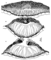
Caption: Fig. 3 Hypoderma liliensis. A-C, ascomata in vertical section at different stages of maturity.
(A, PDD54115; B, PDD 45561; C, PDD 49239). | 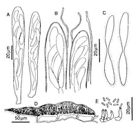
Caption: Fig. 4 Hypoderma liliensis. A, asci. B, apices of asci and paraphyses. C, released ascospores.
D, conidioma in vertical section. E, conidiogenous cells and conidia. (A-C, PDD 46158; D-E,
PDD 57554). | 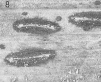
Caption: Fig.8 Macroscopic appearance of ascomata (xl5). Hypoderma liliensis (PDD54788). |
Article: Johnston, P.R. (1991). Rhytismataceae in New Zealand. 4. Pureke zelandicum gen. and sp. nov. plus additional species in Hypoderma, Lophodermium, and Propolis. New Zealand Journal of Botany 29: 395-404 (http://www.rsnz.org/publish/abstracts.php).
Description: Ascomata and conidiomata develop in pale areas on fallen leaves, not associated with zone
lines. Ascomata 1-2.5 x 0.4-0.6 mm, elliptic in outline, walls grey to black, sometimes with a
broad, pale zone developing along the future line of opening shortly before the ascomata open.
Ascomata open by a single longitudinal slit, edge of the opening slit lined with a differentiated
zone, blue-grey in colour when fresh, whitish when dry.
Ascomata subcuticular. In vertical section the ascomatal initial comprises a
lens-shaped group of hyaline, plectenchymatous cells oriented more or less parallel to the surface
of the host leaf, with darkened, angular cells developing toward the lower surface of the
plectenchyma. As the ascomata develop, and paraphyses start to differentiate, darkened
angular cells begin to develop along the upper as well as the lower edge of the ascomata. As
the paraphyses elongate and asci begin to develop the upper wall of the ascomata becomes up
to 60 µm thick near the centre of the ascomata, narrower toward the edges. The upper wall
comprises mostly brown to dark brown, thick-walled, angular cells, but near the centre of the
ascomata, along the future line of opening, toward the inside of the wall, is a group of hyaline
thin-walled, angular cells. The inside of the wall is lined with a poorly developed layer of
hyaline, thin-walled, short-cylindric, downward-oriented cells. In some ascomata the upper
wall retains this appearance until opening, but in others, at about the stage when ascospores
are first starting to differentiate, the cells along the lower part of the pale area along the future
line of opening become darkened and thick-walled. At about the same stage of development,
some ascomata develop an extra layer up to 20 µm thick, of hyaline, thin-walled, vertically
oriented cells between the upper wall and the host cuticle. The upper wall starts to split open
from the outside, and as the wall splits a layer of thin-walled, hyaline, cylindric cells develops
along the exposed face of the broken wall. In opened ascomata the upper wall is up to 100
µm thick near the opening, becoming narrower toward the edge of the ascomata, and
comprises brown to dark brown, thick-walled, angular cells. The exposed face of the broken
upper wall is lined with a palisade-like layer of thin-walled, 10-15 x 1-2 µm, cylindric cells.
The layer between the upper wall and the cuticle present in some ascomata is retained after the
ascomata open. In opened ascomata the lower wall is 5-10 µm thick, comprising 1-3 layers of
brown to dark brown, angular to cylindric cells.
Paraphyses 1.5-2.5 µm diam., undifferentiated at apex, extending 15-25 µm beyond
asci. Asci 130-200 x 18-22 µm, clavate to subsaccate, tapering to truncate apex, wall more or
less undifferentiated at apex, 8-spored, development of asci sequential. Ascospores 40-70 x
5.5-8 µm, bifusiform, 1.5-2.5 µm wide at constriction, 0-1 septate, surrounded by a 2-4 µm
thick gelatinous sheath. Conidiomata 0.2-0.3 mm diam., round in outline, pale brown, with a
darker line around the outside edge, subcuticular. In vertical section the upper wall is more or
less absent. Lower wall 5-10 µm thick, comprising 2-3 layers of brown to dark brown, thick-walled,
angular cells. Conidiogenous cells develop on the lower wall, 6-10 x 2-3 µm, solitary,
cylindric, with percurrent proliferation, wall of the conidiogenous cell thickened and often
slightly flaring at the single, apical conidiogenous locus. Amongst the conidiogenous cells,
near the centre of ascomata, are narrow-cylindric, hyaline, sterile elements extending almost to
the top of the conidiomata. Conidia 4-6 x 1 µm, cylindric, ends rounded, nonseptate, hyaline.
CHARACTERISTICS IN CULTURE: Ascospores germinated from PDD 43973 and 57549
on agar plates, on oatmeal agar colonies 20-30 mm diam. after 5 weeks, surface of colony
undulate, pale brown, aerial mycelium low, felted, white, or lacking, pale brown in reverse.
Habitat: Dead leaves of Astelia nervosa and Collospermum hastatum.
Notes: ETYMOLOGY: refers to the liliaceous host substrate.
NOTES: This is the only species of Rhytismataceae known from Collospermum, but
Coccomyces limitatus, Lophodermium asteliae, and L. minus are known also from Astelia.
Hypoderma liliensis is easily distinguished from these other species by the bluish differentiated
zone along the edge of the ascomatal opening, and microscopically by the large, subsaccate
asci, and bifusiform ascospores.
Although the ascus shape, and ascus and ascospore size, are unusual for Hypoderma, H.
liliensis possesses all the characters used by Johnston (1990) to redefine this genus. Also
atypical of Hypoderma is the darkening of the inner edge of the upper wall seen in some
collections of this species, but as this feature is not found consistently in all collections, it is
not considered of generic significance. The development of a layer of hyaline, cylindric cells
between the upper wall and the cuticle is also unusual. However, a similar phenomenon has
been seen in a few collections of Hypoderma rubi (Persoon:Fries) de Candolle ex Chevallier
from New Zealand, although the layer of cells in this case is not as well developed as in H.
liliensis, and it forms later in the development of the ascomata, appearing only shortly before
the ascomata open.
The size and shape of the asci and ascospores of H. liliensis are similar to those of
Duplicaria empetri (Persoon:Fries) Fuckel and many of the conifer-inhabiting species of
Rhytismataceae. Duplicaria differs in having circular ascomata with radiate opening splits,
and an upper wall with no internal differentiation in unopened ascomata, barely thickened
toward the opening, and with no cylindric, thin-walled cells lining the exposed face of the
broken upper wall in opened ascomata. The conifer-inhabiting species with similar ascospores
and asci differ in having ascomata which develop subepidermally, which lack a darkened lower
wall, and lack differentiated cells along the edge of the opening split. The one conifer-inhabiting genus with large, bifusiform ascospores and subcuticular ascomata, Bifusella, has
synchronous development of the asci, and lacks persistent paraphyses.
|