|
 Hypoderma dundasicum Hypoderma dundasicum
BiostatusPresent in region - Indigenous. Endemic
Images (click to enlarge)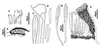
Caption: Fig. 8 Hypoderma dundasicum: A, margin of ascoma in vertical section (PDD48333). B, asci
(PDD 48333). C, apex of asci and paraphyses (PDD 48333). D, released ascospores (PDD
48333). E, conidioma in vertical section (PDD 55170). F, conidiog | 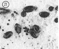
Caption: Fig. 21 Hypoderma dundasicum (PDD 46768). Macroscopic appearance of ascomata (x15). |
Article: Johnston, P.R. (1990). Rhytismataceae in New Zealand 3. The genus Hypoderma. New Zealand Journal of Botany 28(2): 159-183 (http://www.rsnz.org/publish/abstracts.php).
Description: Ascomata and conidiomata developing on fallen leaves, in discrete groups within pale, whitish
areas on host leaves. This paler area often surrounded by narrow, black zone lines. Ascomata
0.6-1.3 x 0.4-0.5 mm, ovate to oblong-elliptic in outline, ends rounded. Unopened ascomata
with uniformly black walls. Opened ascomata with black walls, the single, longitudinal
opening slit lined with a narrow, bright, orange-yellow zone. Conidiomata 0.1-0.2 mm diam.,
round in outline, brown to dark brown, pustulate.
Ascomata subcuticular. In vertical section the upper wall of unopened ascomata is up to
50 µm thick, becoming gradually narrower toward the edge of the ascomata, comprising
mostly brown to dark brown, angular cells, but along the future line of opening, near the
middle of the ascomata, is a wedge-shaped group of paler, thinner-walled cells in the inner
part of the wall. The inside edge of the wall is lined with a few hyaline, cylindric to globose
cells. In opened ascomata the upper wall is 40-55 µm thick, not varying in width in the top
third of the wall, in the lower two-thirds becoming gradually thinner. Wall comprising dark
brown, mostly thick-walled, angular, 5-8 µm diam. cells. Exposed face of the broken upper
wall is lined with hyaline, thin-walled, 15-20 x 3-4 µm, cylindric cells. Lower wall, 10-1 5 µm
thick, of 3-4 layers of dark brown, thick-walled, globose to angular cells.
Paraphyses 1-1.5 µm diam., undifferentiated or loosely circinate at apices, extending 10-15 µm beyond asci. Asci 120-155 x 8-12 µm, clavate-stipitate, tapering to small, truncate
apex, wall undifferentiated at apex, 8-spored, spores confined to upper half of ascus.
Ascospores 21-29 x 2-3 µm (average 23.2 x 2.7 µm), in face view more or less cylindric,
tapering from near the base toward the acute base, 0-septate surrounded by narrow gelatinous
sheath.
Conidiomata subcuticular. In vertical section upper wall 5-10 µm thick, comprising dark
brown material with no visible cellular structure. Lower wall of 1-3 layers of brown to dark
brown, thick-walled, angular cells. Lower wall lined with 1-2 rows of hyaline, thin-walled
cells on which the conidiogenous cells develop. Conidiogenous cells 10-15 x 1.5-2.0 µm,
cylindric, tapering toward apex, with both sympodial and percurrent proliferation, no obvious
thickening of wall at conidiogenous loci. Conidia 3-4.5 µm, straight, cylindric, ends rounded,
hyaline.
Habitat: Fallen leaves of Gaultheria and Pernettya species.
Distribution: Wellington, North Canterbury, MacKenzie, Southland.
Notes: ETYMOLOGY: named after type locality.
NOTES: Hypoderma dundasicum is very similar to the widespread H. rubi. The two species
can be distinguished by ascospore size and shape, and ascus width. The appearance of the
ascomatal wall in vertical section also differs, with the wall of H. rubi decreasing in width
from the edge of the opening, while in H. dundasicum the uppermost part of the wall is more
or less uniform in width. H. rubi has not been found on Ericaceae in New Zealand, although
Powell (1974) reported it on ericaceous hosts from the northern hemisphere. H. gaultheriae
Hunt, described on Gaultheria from North America by Hunt(1980) is reported to be
associated with necrotic spots on green leaves, and has ascospores wider than those of H.
dundasicum.
|