|
 Hypoderma cordylines Hypoderma cordylines
BiostatusPresent in region - Indigenous. Endemic
Images (click to enlarge)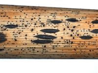
Caption: Ascomata (dry), on Phormium cookianum
Owner: Herb PDD | 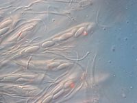
Caption: Ascospores (collection on Phormium cookianum)
Owner: Herb PDD | 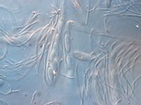
Caption: Ascospores (collection on Phormium cookianum)
Owner: Herb PDD | 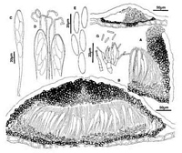
Caption: Fig. 7 Hypoderma cordylines: A, margin of ascoma in vertical section (PDD43225). B,
immature ascoma in vertical section (PDD 53937). C, ascus (PDD 49297). D, apex of asci
and paraphyses (PDD 49297). E, released ascospores (PDD 49297). F, | 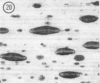
Caption: Fig.20 Hypoderma cordylines (PDD45034). Macroscopic appearance of ascomata (x15). |
Article: Johnston, P.R. (1990). Rhytismataceae in New Zealand 3. The genus Hypoderma. New Zealand Journal of Botany 28(2): 159-183 (http://www.rsnz.org/publish/abstracts.php).
Description: Ascomata and conidiomata developing on dead leaves, within pale, yellowish areas on host
leaf. Pale areas never associated with zone lines. In surface view ascomata 0.8-2.5 x 0.3-0.6
mm, elliptic in outline, tapering to more or less acute ends. Unopened ascomata with grey,
dark grey or black walls, sometimes developing a paler zone along the future line of opening
shortly before the ascomata open. Ascomata opening by a single, longitudinal slit, which is
lined with a narrow, orange to red-brown zone. Conidiomata 0.2-0.3 mm diam., circular in
outline, pale brown with a darker line around the outside edge, pustulate.
Ascomata subcuticular. In vertical section the upper wall of unopened ascomata up to 70
µm thick, narrower toward the edges of the ascomata. Wall comprising mostly brown to dark
brown, angular cells, but with a group of paler, thinner-walled cells in the inner part of the
wall, along the future line of opening. Ascomatal upper wall starts to split open, and a layer of
hyaline, cylindric cells begins to develop along the exposed face of the breaking upper wall,
before covering host cuticle breaks. In opened ascomata the upper wall is up to 70-120 µm
thick near the ascomatal opening, becoming either gradually or more or less abruptly thinner
toward the outside edge. Upper wall comprising dark brown, thick-walled, angular, 4-7 µm
diam. cells. Exposed face of the broken upper wall is lined with hyaline, thin-walled, 20-30 x
4-5 µm, cylindric cells. Lower wall 10-20 µm thick, of 2-3 rows of brown, thick-walled,
angular to cylindric cells.
Paraphyses 1.0-1.5 µm diam., loosely circinate at apex, extending 5-10µm beyond asci.
Asci 90-l40 x 11-16 µm, clavate-stipitate, tapering to truncate apex, wall often slightly
thickened at apex with inconspicuous central pore, 8-spored, spores confined to upper half of
ascus. Ascospores 14-21 x 4.5-6.0 µm (average 17.0 x 5.2 µm), in face view elliptic, tapering
more or less equally to both ends, in side view slightly curved, 0-septate, surrounded by
gelatinous sheath.
Conidiomata subcuticular. In vertical section upper wall absent. Lower wall of 1-3 layers
of brown, thick-walled, angular cells. Lower wall lined with short columns of angular to
cylindric, pale brown to hyaline, 4-8 x 4 µm cells, and the conidiogenous cells develop on this
layer. Conidiogenous cells 13-22 x 2-3 µm, Solitary, cylindric, tapering to apex, with
sympodial proliferation, often with two conidia held at the apex. Conidia 5-9 x 1 µm,
cylindric, straight, ends rounded, 0-septate, hyaline.
CHARACTERISTICS IN CULTURE: Ascospores germinating on agar plates within 24
hours. Colonies on OA 60-80 mm diam. after six weeks, aerial mycelium sparse, white,
cottony, agar surface pale greyish-brown, with numerous, scattered, black-walled, globose
conidiomata. Conidiomata opening by irregular splits in the wall to expose the grey conidial
ooze. Conidiogenous cells and conidia the same as described from plant material.
Habitat: Dead leaves of Cordyline australis, C. banksii, C. indivisa, C. pumilio, less
common on Phormium cookianum and P. tenax.
Distribution: Northland, Auckland, Coromandel, Taupo, Gisbome, Taranaki, Wanganui,
Wellington, Nelson, Buller, North Canterbury.
Notes: ETYMOLOGY: named after host substrate of holotype.
NOTES: Hypoderma cordylines is macroscopically similar to the common and widespread H.
rubi. The two species can be distinguished by ascospore shape, and by the length of the
conidia. H. rubi has not been found on Cordyline spp.
|