|
 Hypoderma cookianum Hypoderma cookianum
BiostatusPresent in region - Indigenous. Endemic
Images (click to enlarge)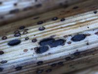
Caption: Ascomata (dry), upper wall sunken slightly below level of surrounding host tissue; single opening slit barely visible.
Owner: Herb PDD | 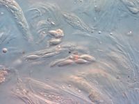
Caption: Ascospores
Owner: Herb PDD | 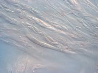
Caption: Ascospores
Owner: Herb PDD | 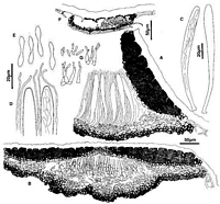
Caption: Fig. 6 Hypoderma cookianum: A, margin of ascoma in vertical section (PDD48455). B,
immature ascoma in vertical section (PDD 54866). C, asci (PDD 48455). D, apex of asci and
paraphyses (PDD 48455). E, released ascospores (PDD 48455). | 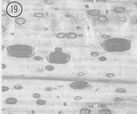
Caption: Fig. 19 Hypoderma cookianum (PDD 54886). Macroscopic appearance of ascomata (x15). |
Article: Johnston, P.R. (1990). Rhytismataceae in New Zealand 3. The genus Hypoderma. New Zealand Journal of Botany 28(2): 159-183 (http://www.rsnz.org/publish/abstracts.php).
Description: Ascomata and conidiomata developing on fallen leaves, within pale, yellowish areas. These
areas often surrounded by narrow, black zone lines. In surface view ascomata 0.8-1.5 x 0.4-0.7 mm, oblong-elliptic in outline, ends broadly rounded. Immature ascomata with black
walls, with a narrow paler zone along the future line of opening, and well developed paler
zones immediately inside the edge of the ascomata, especially toward the ends. Ascomata
opening by a single longitudinal slit, lacking any obvious differentiated cells along the edge of
the opening. Conidiomata circular in outline, 0.2 mm diam., pale brown, darker around
outside edge, immersed.
Ascomata subcuticular. In vertical section upper wall of unopened ascomata up to 45-60
µm thick, narrower toward the outside edge and near the centre of the ascomata. Upper wall
comprising mostly dense, black tissue with no obvious cellular structure, but with brown to
pale brown, angular cells at the narrow, central part of wall. The inside of this central part of
the wall is lined with a few thin-walled, hyaline cells. In opened ascomata the upper wall is up
to 60-90 µm thick, slightly thinner toward the ascomatal opening and toward the edge of the
ascomata. There is no well-differentiated, persistent zone of paler cells lining the exposed face
of the broken upper wall. Lower wall 30-45 µm thick, of 5-10 rows of angular cells, brown to
dark brown and thick-walled toward the outside of the wall, hyaline and thin-walled toward
the inside.
Paraphyses 1.5-2.0 µm diam., undifferentiated at the apex, barely extending beyond asci.
Asci 130-175 x 11-13 µm, clavate, becoming clavate-stipitate immediately prior to spore
release tapering slightly to the broadly truncate to somewhat rounded apex, wall often with a
small pore at the apex, 8spored. Ascospores bifusiform, 22-33 x 3.5-5.0 µm (average 24.5 x
4.1 µm), broadly rounded apex, tapering toward base, constricted to 1.0-1.5 µm diam. near
middle of the spore, surrounded by a narrow gelatinous sheath.
Conidiomata subcuticular. In vertical section the upper wall is 3-5 µm thick, comprising
brown to pale brown material with no obvious cellular structure. Lower wall 5-10 µm thick,
comprising 2-3 rows of angular cells, outer layers of cells brown, inner layers hyaline.
Conidiogenous cells develop on the innermost cells of lower wall, 8-15 x 2-3 µm, cylindric,
solitary, proliferating sympodially, often with two developing conidia at apex. Conidia 4-6 x I
µm, cylindric, ends rounded, straight, 0-septate, hyaline.
Habitat: Dead leaves of Phormium cookianum.
Distribution: Coromandel, North Canterbury.
Notes: ETYMOLOGY: named after host substrate.
NOTES: Ascomatal shape and ascospore shape distinguish this species from H. cordylines,
also found on Phormium in New Zealand.
See also notes under H. carinatum.
|