|
 Hypoderma carinatum Hypoderma carinatum
BiostatusPresent in region - Indigenous. Endemic
Images (click to enlarge)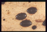
Owner: P.R. Johnston | 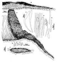
Caption: Fig.5 Hypoderma carinatum: A, margin of ascoma in vertical section (PDD49290). B,
immature ascoma in vertical section (PDD 49291). C, asci (PDD 49290). D, apex of asci and
paraphyses (PDD 49290). E, released ascospores (PDD 49290). F, coni | 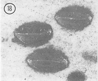
Caption: Fig. 18 Hypoderma carinatum (PDD 46709). Macroscopic appearance of ascomata (x15). |
Article: Johnston, P.R. (1990). Rhytismataceae in New Zealand 3. The genus Hypoderma. New Zealand Journal of Botany 28(2): 159-183 (http://www.rsnz.org/publish/abstracts.php).
Description: Ascomata and conidiomata developing on fallen leaves, in discrete groups within pale,
yellowish areas on host leaf. This paler area often surrounded by narrow, black zone line. In
surface view ascomata 1.8-3.0 x 1.0-1.7 mm, broad-elliptic in outline with rounded ends.
Unopened ascomata with uniformly black walls, with host tissue immediately surrounding the
ascomata often stained rusty brown. Opened ascomata forming a keel-like ridge along the
single, longitudinal opening slit, wall not otherwise differentiated along opening. Conidiomata
0.2-0.3 mm diam., circular in outline, brown to dark brown becoming almost black with age,
pustulate.
Ascomata subcuticular. In vertical section the upper wall of unopened ascomata up to
30-40 µm thick, narrower toward edges and at the centre of the ascomata. Upper wall
comprising mostly dense, black tissue with no obvious cellular structure, except for the inner
part of the wall along the future line of opening, where it comprises thin-walled, hyaline cells.
In opened ascomata the upper wall is up to 60-80 µm thick, becoming gradually thinner
toward the base of the wall, and toward the ascomatal opening. A few poorly developed,
hyaline, cylindric cells initially line the exposed face of the broken upper wall, but these cells
are not persistent. Lower wall 5-10 µm thick, of 1-2 layers of brown to pale brown, angular
to cylindric cells. Upper and lower walls extending up to 300-400 µm beyond hymenium.
Paraphyses 1.0-3.5 µm diam., undifferentiated or slightly and irregularly swollen near
apices, extending 5-10 µm beyond asci. Asci 110-180 x 10-12 lam, clavate-stipitate, tapering
gradually to small, truncate apex, wall undifferentiated at apex, 8-spored, spores restricted to
upper half of ascus. Ascospores 32-45 x 2.5-4.5 µm (average 37.9 x 3.0 µm), in face view
cylindric, tapering slightly to base, with slight or sometimes well-developed constriction near
centre of spore, 0-1-septate, surrounded by gelatinous sheath. In many asci ascoconidia are
produced from both ends of unreleased ascospores. Asci often becoming packed with the 2.5-6.0 x 1 µm, cylindric, hyaline, 0septate ascoconidia.
Conidiomata subcuticular. In vertical section lenticular in shape, upper wall 5 µm thick,
comprising dense, brown tissue with no visible cellular structure. Lower wall of 1-2 layers of
cylindric to angular, brown to dark brown, thick-walled cells. Lower wall lined with 1-2
layers of thin-walled, hyaline cells on which the conidiogenous cells develop. Conidiogenous
cells 6-11 x 1.5-2.5 µm, cylindric, percurrent proliferation, wall thickened, and sometimes
flaring, at the single, apical conidiogenous locus. Conidia 3.0-5.0 x 1.0-1.5 µm, cylindric,
straight, ends rounded, 0-septate, hyaline.
CHARACTERISTICS IN CULTURE: Ascospores from PDD 46709 germinated within 24
hours on agar plates. Ascoconidia did not germinate. Cultures on OA 60 mm diam. after 8
weeks, aerial mycelium white, coarse, cottony, tufted, agar surface grey-green to olivaceous
with black patches. Scattered, globose, black-walled conidiomata, conidiogenous cells as
described from host material, conidia longer, 5-7 x 1-1.5 µm.
Habitat: Dead leaves of Dracophyllum traversii.
Distribution: Coromandel, Nelson, Buller, Westland.
Notes: ETYMOLOGY: carinatum = keeled; refers to ascomatal shape, with keel-like ridge along
opening slit.
NOTES: H. carinatum is easily confused with two other species with similarly sized asci and
ascospores, H. campanulatum and H. alborubrum. H. carinatum can be distinguished by its
very broad ascomata, greater than I mm wide, and by the keel-like ridge along the ascomatal
opening.
The appearance in vertical section of the upper wall of unopened ascomata of H.
carinatum is similar to that of H. cookianum. Both species have an ascomatal upper wall
thinner along the future line of opening, and with no persistent layer of differentiated cells
along the edge of the ascomatal opening. As in other Hypoderma species the ascomata are
subcuticular, and the inner part of the upper wall along the future line of opening comprises
cells thinner-walled and paler than those of the rest of the wall, the paraphyses are
undifferentiated at the apex, and the asci are clavate-stipitate.
See also notes under H. campanulatum, H. alborubrum.
|