|
 Hypoderma campanulatum Hypoderma campanulatum
BiostatusPresent in region - Indigenous. Endemic
Images (click to enlarge)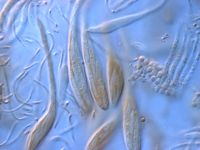
Caption: Ascospores
Owner: Herb PDD | 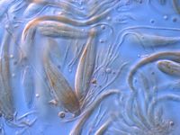
Caption: Ascospores
Owner: Herb PDD | 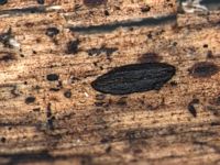
Caption: Ascoma (dry)
Owner: Herb PDD | 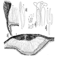
Caption: Fig. 4 Hypoderma campanulatum: A, margin of ascoma in vertical section (PDD 49288). B,
immature ascoma in vertical section (PDD 49283). C, asci (PDD 49283). D, apex of asci and
paraphyses (PDD 49283). E, released ascospores (PDD 49283). F, | 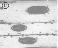
Caption: Fig.17 Hypoderma campanulatum (PDD 54116). Macroscopic appearance of ascomata (x15). |
Article: Johnston, P.R. (1990). Rhytismataceae in New Zealand 3. The genus Hypoderma. New Zealand Journal of Botany 28(2): 159-183 (http://www.rsnz.org/publish/abstracts.php).
Description: Ascomata and conidiomata developing on fallen leaves, in discrete groups within pale,
yellowish areas on host leaf. Pale areas often surrounded by narrow, black zone lines.
Ascomata 1.5-3.0 x 0.5-1.0 mm, elliptic in outline with rounded ends. Ascomata mostly
black-walled, but paler, and grey near the outside edge. This paler area is flattened, forming a
shelf-like margin around the otherwise raised ascomata. Ascomata open by a single,
longitudinal slit, lined with an often indistinct yellow, brown, or dark brown zone.
Conidiomata 0.2-0.3 mm diam., circular in outline, brown to dark brown, pustulate.
Ascomata subcuticular. In vertical section, upper wall of the unopened ascomata up to
60-70 µm thick, comprising mostly brown to dark brown, angular cells, with darker areas
along the outer and inside edges of wall, but with a line of paler, thin-walled cells at the centre
of the ascomata, along the future line of opening. In opened ascomata the upper wall is up to
90-1 1 0 µm thick near the ascomatal opening, becoming abruptly thinner about half way to
the edge of the ascomata. Upper wall comprising either all dense, black tissue with no visible
cellular structure, or with a few thick-walled, dark brown, angular cells visible in the centre of
the labia. Exposed face of the broken upper wall lined with hyaline to pale brown, thin-walled, cylindric cells. Lower wall 3-5 µm thick, of 1-2 layers of thick-walled, pale brown,
angular, 3-5 µm diam. cells. Both walls extending over 200 µm beyond edge of hymenium.
Paraphyses 1-1.5 µm diam., circinate at apices, extending 5-10 µm beyond asci. Asci
110-175 x 11-14 µm, clavate-stipitate, tapering to small, truncate apex, wall undifferentiated
at apex, 8-spored, spores confined to upper half of ascus. Ascospores 28-45 x 2.5-3.5 µm
(average 37.2 x 2.9 µm), in face view cylindric, apex rounded, near the base tapering to more
or less acute base, 0-septate, surrounded by gelatinous sheath.
Conidiomata subcuticular. In vertical section lenticular in shape, upper wall 5 µm thick,
of brown to black material with no visible cellular structure. Lower wall of 2-3 layers of
brown to dark brown, thick-walled, angular cells. Lower wall is lined with 1-2 layers of thin-walled, hyaline cells on which the conidiogenous cells develop. Conidiogenous cells 6-9 x
2.0-2.5 µm, cylindric, percurrent proliferation, wall thickened at single apical conidiogenous
locus. Conidia 3-4 x 1 µm, cylindric, straight, 0-septate, hyaline.
Habitat: Dead leaves of Dracophyllum latifolium and D. traversii.
Distribution: Northland, Auckland, Coromandel, Gisborne.
Notes: ETYMOLOGY: campanulatus = bell-shaped; refers to ascomatal shape, with a distinctive,
flattened, shelf-like plate of tissue surrounding the central, raised part of the ascomata.
NOTES: H. campanulatum is often found in association with two other species, H.
carinatum and H. alborubrum. The most distinctive feature of H. campanulatum is the paler,
grey, flattened, shelf-like margin to the ascomata.
See also notes under H. carinatum and H. alborubrum.
|