|
 Hypoderma bidwillii Hypoderma bidwillii
BiostatusPresent in region - Indigenous. Endemic
Images (click to enlarge)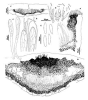
Caption: Fig. 2 Hypoderma bidwillii: A, margin of ascoma in vertical section (PDD53902). B,
immature ascoma in vertical section (PDD 53902). C, asci (PDD 49130). D, apex of asci and
paraphyses (PDD 49130). E, released ascospores (PDD 49130). F, con | 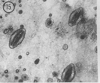
Caption: Fig.15 Hypoderma bidwillii (PDD53902). Macroscopic appearance of ascomata (x15). |
Article: Johnston, P.R. (1990). Rhytismataceae in New Zealand 3. The genus Hypoderma. New Zealand Journal of Botany 28(2): 159-183 (http://www.rsnz.org/publish/abstracts.php).
Description: Ascomata and conidiomata developing in discrete groups on upper surface of fallen leaves.
Not associated with pale areas on leaf, or zone lines. In surface view ascomata 0.8-2.0 x 0.5-0.6 mm, elliptic in outline, acute to both ends. Unopened ascomata with pale grey walls, with
broad, paler zone along the future line of opening. Near centre of the ascomata the pale zone
is interrupted by a small, circular dark patch, which extends about half way toward sides of the
ascomata. Outside edge of the ascomata is marked by a narrow, dark line. Ascomata open by
a single, longitudinal slit, the edge of which is lined with a broad, pale pinkish-brown zone.
The dark patch visible at the centre of immature ascomata is still evident in mature ascomata,
but is divided in half by the opening slit. Conidiomata circular in outline,0.1-0.2mm diam.,
pale brown, darker around outside edge, immersed.
Ascomata subcuticular. In vertical section the upper wall of unopened ascomata is up to
70 µm thick, comprising mostly brown to dark brown, thick-walled, angular cells. The outer
part of the upper wall near the centre of the ascomata is composed of dense, black tissue with
no obvious cellular structure, and the dense, black material often extends into what appear to
be cracks in the host cuticle. The inside of this part of the upper wall is lined with a layer of
hyaline, somewhat disorganised tissue. A narrow extension to this hyaline layer extends
vertically through part of the dense, black layer in the outer part of the wall. In opened
ascomata the upper wall is up to 85 µm thick near the ascomatal opening, becoming abruptly
thinner, to 10-25 µm thick in lower half. Wall comprising brown to dark brown, thick-walled
5-7 µm diam. cells, with dense, black tissue with no obvious cellular structure in the outer part
of the wall, near opening slit. Exposed face of the broken upper wall is lined with a layer of 1-4 µm diam., branching, hyaline, thin-walled, cylindric cells. Lower wall 20-40 µm thick, of
angular cells, cells dark brown and thick-walled in outer part of wall, paler and thinner-walled
in inner part.
Paraphyses 0.8-1.0 µm diam., apices undifferentiated or loosely circinate, extending 20-30 µm beyond asci. Asci 140-160 x 12-17 µm, clavate-stipitate, tapering to small, truncate
apex, wall thinner at apex, 8-spored, spores confined to upper half of ascus. Ascospores 21-36 x 4.5-5.5 µm (average 24.8 x 4.8 µm), in face view oblong-elliptic, tapering slightly to
rounded ends, 0-septate, surrounded by narrow, gelatinous sheath. Conidiomata subcuticular.
In vertical section the upper wall is absent. The lower wall is 5-10 µm thick, of 1-2 rows of
thick-walled, brown, angular to cylindric cells. Conidiogenous cells solitary or on short
conidiophores, developing on the lower wall, 10-18 x 2-3 µm, cylindric, with percurrent
proliferation, sometimes with several distinct annelations near apex. Conidia 5-10 x 1 µm,
cylindric, straight, ends rounded, 0-septate, hyaline.
Habitat: Fallen leaves of Brachyglottis bidwillii.
Distribution: Taupo, Wellington.
Notes: ETYMOLOGY: named after host substrate.
Hypoderma bidwillii can be distinguished by macroscopic appearance. The
immature ascomata have a distinctive paler zone along the future line of opening, with this
paler zone interrupted near the centre of the ascomata by a small, circular patch of dark tissue.
In addition, the distribution of dense, black tissue in the ascomatal upper wall differs from that
seen in all other New Zealand Hypoderma spp.
|