|
 Hypoderma alborubrum Hypoderma alborubrum
BiostatusPresent in region - Indigenous. Endemic
Images (click to enlarge)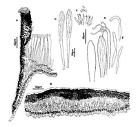
Caption: Fig. 1 Hypoderma alborubrum: A, margin of ascoma in vertical section (PDD 53984). B,
immature ascoma in vertical section (PDD 49286). C, asci (PDD 53984). D, apex of asci and
paraphyses (PDD 53984). E, released ascospores (PDD 53984). F, c | 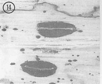
Caption: Fig.14 Hypoderma alborubrum (PDD53984). Macroscopic appearance of ascomata (x15). |
Article: Johnston, P.R. (1990). Rhytismataceae in New Zealand 3. The genus Hypoderma. New Zealand Journal of Botany 28(2): 159-183 (http://www.rsnz.org/publish/abstracts.php).
Description: Ascomata and conidiomata developing on fallen leaves, in discrete groups within pale,
yellowish areas of host leaf. Pale area often surrounded by narrow, black zone line. In surface
view ascomata 1.2-3.0 x 0.5-1.0 mm, elliptic in outline, often acute to the ends. Unopened
ascomata uniformly black-walled, edge usually sharp, sometimes with black flecking in
surrounding host tissue. Ascomata opening by single longitudinal slit, which is lined with a
narrow, white or bright red zone. Conidiomata 0.2-0.3 mm diam., round in outline, brown to
dark brown, pustulate.
Ascomata subcuticular. In vertical section upper wall of unopened ascomata up to 50-60
µm thick. Inner and outer parts of the wall comprise dense, black tissue with no obvious
cellular structure, otherwise mostly of dark brown, angular cells. Along the future line of
opening, in the inner part of the wall, is a group of thinner-walled, paler cells. In opened
ascomata the upper wall is 50-70 µm thick near the opening slit, becoming abruptly thinner
about half way to the base of the wall, comprising mostly brown to pale brown, thick-walled,
angular cells, but with a patch of dense, black tissue on the inner edge of the wall near the
ascomatal opening, and along outermost 5-10 lam of the wall. Exposed face of the broken
upper wall is lined with a compact layer of cylindric, hyaline, thin-walled cells. Lower wall 5
µm thick, of 1-2 layers of brown to pale brown, angular cells.
Paraphyses 1.0-1.5 µm diam., circinate at apices, extending 10-15 µm beyond asci. Asci
120-160 x 11-14 µm, clavate-stipitate, tapering to small, truncate apex, sometimes with tiny
apical pore, 8spored, spores confined to upper half of ascus. Ascospores 35-45 x 3-4 µm
(average 41.2 x 3.1 µm), in face view cylindrical-bifusiform, rounded at apex, tapering near
base, 1-1.5 µm wide at central constriction, 0-septate, surrounded by gelatinous sheath.
Conidiomata subcuticular. In vertical section lenticular in shape, upper wall 5 µm thick,
of brown material with no visible cellular structure. Lower wall of 2-3 layers of brown to dark
brown, thick-walled, angular cells. Lower wall is lined with hyaline, thin-walled cells, on
which the conidiogenous cells are held. Conidiogenous cells 7-20 x 2-3 µm, flask-shaped,
with a broad base and narrow apex, sympodial proliferation, often with more than one
conidium held at apex. Conidia 3-4 x 1 µm, cylindric, straight, 0-septate, hyaline.
Habitat: Dead leaves of Dracophyllum menziesii and D. traversii.
Distribution: Gisborne, Nelson, Buller.
Notes: ETYMOLOGY: albo = white, rubrum = red; refers to the white or bright red zone of cells
lining the ascomatal opening.
H. alborubrum can be difficult to distinguish from two other species often found on
the same leaves, H. campanulatum and H. carinatum. The most distinctive macroscopic
feature of H. alborubrum is the zone of bright, white or red cells lining the opening slit.
Microscopically this species has distinctly bifusiform ascospores, while the other two species
have ascospores which are only slightly, if at all, constricted. The anamorph of H.
alborubrum also differs in having conidiogenous cells which proliferate sympodially rather
than percurrently, and in lacking any wall thickening at the conidiogenous locus.
See also notes under H. carinatum and H. campanulatum.
|