|
 Gymnomyces redolens Gymnomyces redolens
SynonymsOctaviania redolens
Martellia redolens
Stephanospora redolens
BiostatusPresent in region - Indigenous. Endemic
Images (click to enlarge)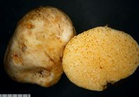
Owner: J.A. Cooper | 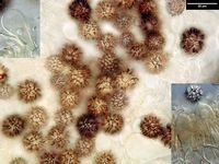
Owner: J.A. Cooper | 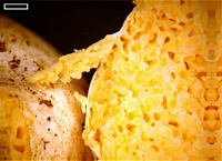
Caption: scale=1mm
Owner: J.A. Cooper | 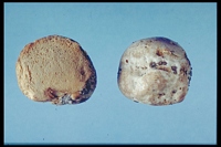
Owner: R.E. Beever | 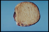
Owner: R.E. Beever | 
Owner: R.E. Beever | 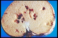
Owner: R.E. Beever | 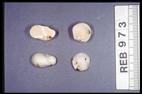
Owner: R.E. Beever | 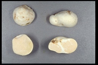
Owner: R.E. Beever | 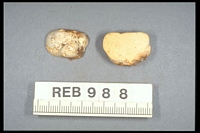
Owner: R.E. Beever | 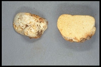
Owner: R.E. Beever | 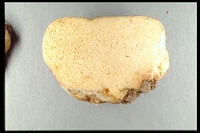
Owner: R.E. Beever | 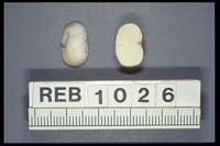
Owner: R.E. Beever | 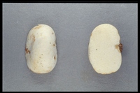
Owner: R.E. Beever | 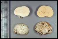
Owner: R.E. Beever | 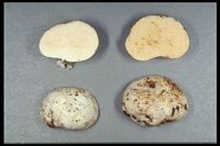
Owner: R.E. Beever | 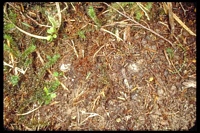
Owner: R.E. Beever | 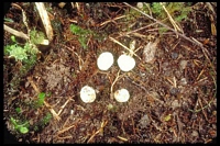
Owner: R.E. Beever | 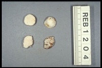
Caption: herb. REB 1204
Owner: R.E. Beever | 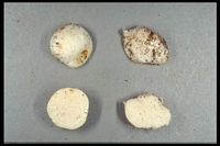
Caption: herb. REB 1204
Owner: R.E. Beever | 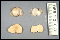
Caption: herb. REB 1208
Owner: R.E. Beever | 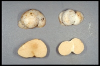
Caption: herb. REB 1208
Owner: R.E. Beever | 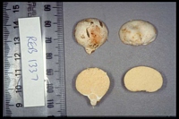
Owner: R.E. Beever | 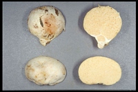
Owner: R.E. Beever | 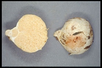
Owner: R.E. Beever | 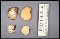
Caption: [specimen in photograph labelled in error as REB 1434]
Owner: R.E. Beever | 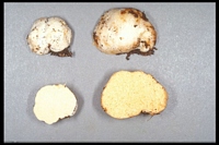
Owner: R.E. Beever | 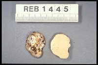
Owner: R.E. Beever | 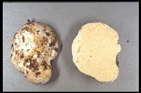
Owner: R.E. Beever | 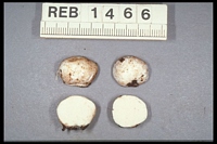
Owner: R.E. Beever | 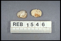
Owner: R.E. Beever | 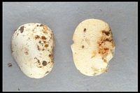
Owner: R.E. Beever | 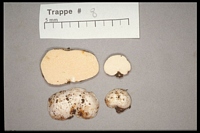
Owner: R.E. Beever | 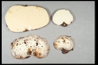
Owner: R.E. Beever | 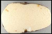
Owner: R.E. Beever | 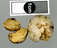
Owner: J.A. Cooper | 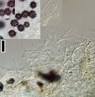
Caption: scale=20um. Upper spores and basidia. Lower cutis.
Owner: J.A. Cooper | 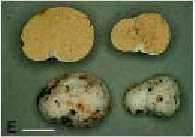
Caption: Fig. 3 E, Gymnomyces redolens; Scale bar = 10 mm. | 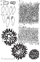
Caption: Fig. 10 Gymnomyces redolens. A, Basidioma; B, peridiopellis and peridial context; C,
hymenophoral trama and hymenium; D, basidia; E, hymenophor | 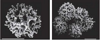
Caption: Fig. 11 Spores of Gymnomyces redolens. Scale bars = 10 mm. | 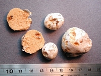
Caption: fruitbody
Owner: J.A. Cooper | 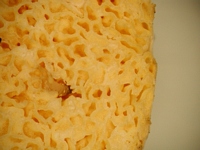
Caption: fruitbody
Owner: J.A. Cooper | 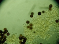
Caption: spores and basidia in Melzer's
Owner: J.A. Cooper | 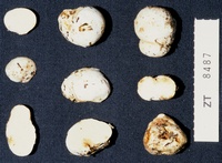
Caption: ZT8487
Owner: E. Horak: © Creative Commons Attribution-Noncommercial 3.0 New Zealand | 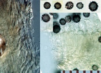
Caption: left: section through cuticle. Right basidia and stalked spores (melzers)
Owner: J.A. Cooper | 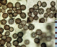
Caption: spores (melzers)
Owner: J.A. Cooper | 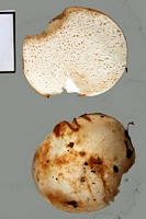
Owner: J.A. Cooper | 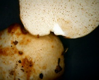
Owner: J.A. Cooper | 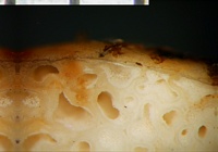
Owner: J.A. Cooper | 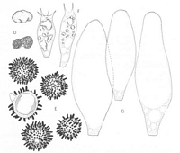
Caption: FIG. 4. D-G. Martellia redolens: D. habit and section. x 1 E. spores. x 1750
F. basidia. x 1000 G. cystidia. | 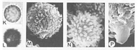
Caption: Plate 25. K-P. Martellia redolens (Beaton 27): K-L. light micrographs. x 1250; K.
unstained, L. stained M. lateral. SEM x 3250 N. apical. SEM x 3000 P. hilar appendix. SEM x 5000 | 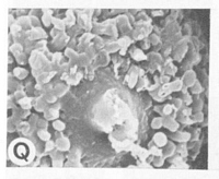
Caption: Plate 25 Q. Martellia redolens (Beaton 27): base. SEM x 4000 | 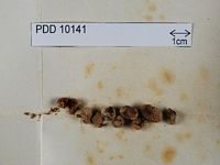
Caption: Dried type specimen
Owner: Herb PDD | 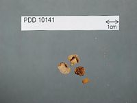
Caption: Dried type specimen
Owner: Herb PDD |
Article: Lebel, T. (2002). Sequestrate Russulales of New Zealand: Gymnomyces and Macowanites. New Zealand Journal of Botany 40(3): 489-509 (http://www.rsnz.org/publish/abstracts.php).
Description: Basidiomata
5-25 mm diam., subglobose or irregular, depressed around basal attachment, rarely with an
exposed gleba. Peridial surface slightly tomentose, finely wrinkled or smooth, white to cream-coloured
drying pallid ochraceous. Context thin, fragile, off-white. Gleba white to
cream becoming pallid ochraceous, loculate, locules small,irregular. Stipe absent. Columella
absent or present, rudimentary, percurrent. Odour when fresh pleasant, resembling dried
apricots; taste not recorded. Latex absent. Peridiopellis 70-150 µm wide, a dense turf of
upright to repent, hyaline hyphal tips 20-55 x 2-4.5 µm diam., becoming tangled and
interwoven. Peridial context 210-450 µm wide, of tightly interwoven, non gelatinised, hyaline
hyphae 2-3.5 µm diam., sphaerocysts 12-22 µm diam. in scattered nests. Endocystidia absent.
Columella context when present of interwoven, hyaline hyphae 2-3 µm diam. Hymenophoral
trama 40-75 µm wide,of interwoven, hyaline hyphae 2-4.5 µm diam., not gelatinised, with
sphaerocysts 12-27 µm diam. in scattered nests. Subhymenium poorly developed, 11-20 µm
wide, with 1-2 tiers of isodiametric cells 5-9 µm diam. Basidia 25-35 x 9-11 µm,
hyaline, ventricose to clavate, mostly with 1 or rarely 2 sterigmata 3-5 x 1-2 µm, robust.
Cystidia 24-49 x 8-15 µm, broadly ventricose with broadly rounded apices and few granular
contents refractive in KOH; arising in subhymenium, not extending much beyond basidia, rare.
Spores 9-12.5(-14) x 9-12(-14) µm (10.88 ± 0.32 x 10.09 ± 0.3), Q = 1.05-1.07, globose to
subglobose, orthotropic and symmetric, wall hyaline. Ornamentation amyloid, a dense
spiny reticulum of warts and spines 2-4 µm high, bases just coalescing or several joined at
their bases by low lines 0.2-0.8 µm high or ridges ± 1-1.5 µm high in a partial reticulum. Hilar
appendix 1-1.5 x 0.5-1 µm, cylindrical; plage inamyloid. Spore colour in mass hyaline, in the
locules of the dried gleba appearing pale cream coloured.
Habitat: HABITAT AND DISTRIBUTION: Hypogeous, growing in scattered groups among leaf litter
in Nothofagusfusca, N. solandrii, and N. menziesii forests andLeptospermum scoparium or
Kunzea ericoides scrub. Fruiting period Mar-Nov.
Notes: NOTES: Gymnomyces redolens is widely distributed in New Zealand. It differs from other
New Zealand taxa in the robust spiny-reticulate ornamentation of the spores, and the mono- or
bi-sterigmate basidia.The nomenclatural history of this species is given in Lebel & Castellano
(2002).
Article: Cunningham, G.H. (1942). Two additional New Zealand Gasteromycetes. New Zealand Journal of Science and Technology. B. General Section 23: 172-173.
Description: Plants subglobose or tuberiform, white or cream coloured; becoming pallid ochraceous, 0.5-2.5 cm.
diameter. Peridium exteriorly slightly tomentose, finely wrinkled or smooth, to 450 µ
thick, composed of a single layer of hyaline, woven, non-gelatinized hyphae. Gleba pallid
ochraceous, cells compressed, two or more to millimetre, empty; sterile base absent; tramal
plates 75 x 100 µ thick, of woven hyphae, not gelatinized, fragile, scissile, especially at the
gussets; basidia one-spored. Spores globose or subglobose, 14-22 µ diameter (including
spines), apedicellate, epispore hyaline, 2 µ thick, closely covered with narrowly wedge-shaped spines,
acuminately pointed, basally often merging and giving to the spore a
reticulated appearance, to 4.5 µ long and hyaline.
Notes: When freshly collected the species had a pleasant fragrant smell resembling dried apricots. It
is separated from others of the genus present in New Zealand by the single woven layer of the
peridium, monosporous basidia and large spores with thick epispore and strongly developed
echinulations. Specimens were collected among debris on the forest floor adjoining tracks in
rain forest at the base of Mt. Te Aroha.
Article: Beaton, G.W.; Pegler, D.N.; Young, T.W.K. (1984). Gasteroid Basidiomycota of Victoria State, Australia. 2. Russulales. Kew Bulletin 39(4): 669–698.
Description: Gasterocarp 0.5-2.5 cm diam., subglobose, ellipsoid or irregularly turbinate, depressed
around the basal attachment, very occasionally with an exposed gleba. Peridium white when
fresh, drying greyish orange, farinose to sub-tomentose, smooth finally wrinkled. Gleba
white, cream coloured or pale ochraceous, labyrinthoid, of minute, elongated or irregular,
empty chambers, 2-4 per mm, with some radial and concentric arrangement. Tramal plates
very thin, 75-100 µm thick, consisting of a narrow hymenophoral trama, lacking
sphaerocytes, and moderately well developed subhymenial layers. Columella absent or
sometimes poorly developed; sterile base minute. Latex and laticiferous elements absent.
Spore deposit pale cream coloured. Spores 7-10 x 7-9(9 ± 0.4 x 8-5 ± 0-3) µm (excl. orn.), Q=
1.05; orthotropic, globose or nearly so, hyaline, thin-walled, with an ornamentation of hollow,
tapering spines, with rounded apices, often coalescing at their bases but without connectives,
strongly amyloid; hilar appendix short, 1-1.5 x 0.5-1 µm, cylindric or obconical, with a
terminal hilar tear. Basidia 25-37 x 9-11 µm, ventricose-clavate, bearing 2 or 4 slender
sterigmata. Leptocystidia present, voluminous, 50-80 x 17-35 µm, ovoid-pedicellate to short
lageniform with a broadly rounded apex, and few contents. Hymenophoral trama narrow,
more or less regular, of parallel, filamentous hyphae, 2.5-6.5 µm diam. Subhymenial layer 11-16 µm
broad, pseudoparenchymatous. Peridiopellis a stratified epithelium, up to 200 µm
thick, of agglutinated sphaerocytes, 15-40 µm diam. (Fig. 4 D-G, Pl. 25 K-Q).
Notes: A small hypogeal species, growing in scattered groups, which was originally described from
New Zealand. The amyloid, orthotropic spores place this species in Elasmomycetaceae,
whilst the absence of sphaerocytes in the hymenophoral trama, together with the spinose
spore ornamentation, and the voluminous leptocystidia indicate Martellia to be the most
appropriate genus.
|