|
 Tubaria lanatula Tubaria lanatula
SynonymsPhaeomarasmius lanatulus
BiostatusPresent in region - Indigenous. Endemic
Images (click to enlarge) | 
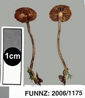
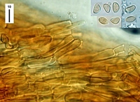
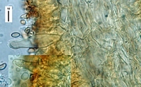
Caption: FUNNZ: 2006/1175, See public note for more information
Owner: FUNNZ | 
Caption: FUNNZ: 2006/1175, See public note for more information
Owner: FUNNZ | 
Caption: FUNNZ: 2006/1175, See public note for more information
Owner: FUNNZ | 
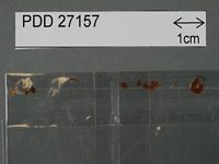
Caption: Dried type specimen
Owner: Herb PDD |
Article: Horak, E. (1980). Fungi Agaricini Novazelandiae. VIII. Phaeomarasmius Scherffel and Flammulaster Earle. New Zealand Journal of Botany 18(2): 173–182 (http://www.rsnz.org/publish/abstracts.php).
Description: Pileus -10mm diam., hemispheric when young, becoming convex and finally plane to expanded; whitish to pale brown, densely covered with conspicuous, woolly or felty brown to black-brown fibrils and squamules (especially at disc); dry, veil remnants on non-striate margin absent. Lamellae (L 8-12, -3) moderately crowded, broadly adnate to emarginate; whitish at first, turning cinnamon-; brown or pale argillaceous, edge albofimbriate. Stipe -15 x-1 mm, cylindric, central, equal; concolorous with pileus, covered with brown woolly fibrils, apex pruinose; dry, hollow, single in groups, distinct veil remnants lacking. Context pale brown. Odour none. Spores 8-10 x 4-5 µm, amygdaliform to sublimoniform, mucro distinct, brown, smooth, thin-walled, germ pore absent. Basidia 20-30 x 5-6µm, 4-spored. Cheilocystidia 35-50 x 8-13 µm, up to; 11 µm at apex, fusoid with distinct capitate neck, membrane thin-walled, occasionally with brown plasmatic pigment, clamp connection at basalt septum, crowded on lamellar edge. Pleurocystidia absent. Cuticle a trichoderm of bundled cylindric hyphae (3-7 µm diam.), terminal cells not differentiated, membranes thin-walled, not gelatinised encrusted with brown (KOH) pigment. Clamp connections numerous.
Habitat: on rotten wood and bark of Nothofagus fusca (Hook.f.) Oerst. New Zealand.
Notes: This delicate agaric is common on well rotten logs and stumps of Nothofagus fusca (red beech) Macroscopically P. lanatulus resembles most P. hispidulus Horak from New Zealand and P. nanus (Horak 1979) from Papua New Guinea. However, these two morphologically similar taxa are well separated by the size and shape of the spores.
|