|
 Dicephalospora chrysotricha Dicephalospora chrysotricha
SynonymsChlorosplenium chrysotrichum
Peziza chrysotricha
Trichopeziza chrysotricha
BiostatusPresent in region - Indigenous. Endemic
Images (click to enlarge)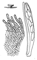
Caption: FIG. 29. Chlorosplenium chrysotrichum. Habit sketch x 8, details x 660. | 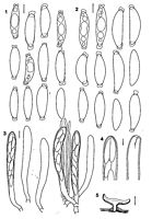
Caption: Dicephalospora chrysotricha (in water). 1, 2- Ascospores. -3. Asci and paraphyses. -4. Ascus tips before and after dehiscence. -5. Section through apothecium. Scale bars: 1, 2, 4= 5 µm, 3= 10 µm; 5= 0.5 mm. -1, 3, 5, from PDD 78708, G.V. 1886, 2 from PDD | 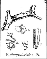
Caption: PLATE CV. Fig. 7. Peziza chrysotricha, natural size. a, hairs; b, external cells; c, ascus and
paraphyses - magnified. d. Sporidia, magnified 250 diameters. |
Article: Dennis, R.W.G. (1961). Some inoperculate Discomycetes from New Zealand. Kew Bulletin 15(2): 293-320.
Notes: This beautiful species was fully described by Massee (in Journ. Linn. Soc. Bot. 31: 517
(1896)), but its systematic position still presents some difficulty. It is not a Trichopeziza as
typified by Saccardo but it agrees tolerably in structure with the species of Chlorosplenium de
Not. The asci, however, give a negative pore reaction with iodine and their spores are an odd
shape for that genus.
Article: Berkeley, M.J. (1855). Fungi. Flora Novae-Zelandiae. Part II. Flowerless Plants. The Botany of the Antarctic Voyage II Hooker, J.D. (eds.) 378 p. 172-210, 338 London: Lovell Reeve.
Description: About a line across. Cups bright golden-yellow, at first globose, then subhemispherical,
sometimes with a slight membranous edge, thickly clothed with short, matted hairs,
somewhat pulverulent. Hymenium closed when dry, seated on a stratum of large, but
unequary-sized cells. Asci cylindrical, rather large; sporidia cymbiform, slightly attenuated at
either end, 1/2250 of an inch long.
Habitat: On twigs on the shores of Waikare Lake, Colenso.
Notes: A remarkably beautiful species.
Article: Saccardo, P.A. (1889). Discomyceteae et Phymatosphaeriaceae. Sylloge Fungorum 8: 3-859 Padua: (http://194.203.77.76/LibriFungorum/Search.asp?ItemType=I).
Habitat: in ramis pr. Waikare Lake Novae Zelandiae (COLENSO).
Notes: Ascoma 2-3 mm. lat.
Article: Verkley, G.J.M. (2004). Redisposition of Chlorosplenium chrysotrichum to the genus Dicephalospora (Sclerotiniaceae, Ascomycota). Sydowia 56(2): 343-348.
Description: Comb. Nov.
Apothecia scattered, erumpent from stromatized inner layers of the bark of the host. - Disc concave or flat, circular, smooth, pale orange-yellow and up to 5 mm diam when moistened, and orange to orange-brown, 1-3.5 mm diam and laterally enfolded when dry. Receptacle cupulate, relatively thin, the surface clothed with hair-like hyphal extensions, intensely orange when dry. Stipe central, relatively thin, orange, somewhat darker or black towards the base. Ectal excipulum 25-50 µm thick, golden yellow to orange, in the upper-part of the receptacle composed of isodiametric, irregular cells 5-10 um diam, downwards transformed into septate, undulating hyphae with hyaline, refractive walls thickened up to 2 µm and enclosing golden yellow particles, the outermost cells also giving rise to thin, tightly appressed, septate hair-like hyphae 20-50x3.5-5 µm, with orange incrustrations on the surface. Medullary excipulum up to 200 µm thick near the stipe, hyaline to pale yellow, almost entirely composed of tightly interwoven, hyaline hyphae 2.5-4 µm wide, in the lower part with somewhat thickened walls. Asci 8-spored, cylindrical-clavate, apex conical-rounded, the walls thickened up to 1 µm, the wall in the top somewhat thinner, not blueing in iodine (IKI-, Mlz-), opening by an irregular tear at the apex, gradually narrowed towards the base, croziers present, 95-120(-130) x 10-14 µm (NT, water; in the type 90-100 x 11-12 µm cf. Massee, 1896: 517; 75-96 x8-12 µm cf. Dixon, 1974). Ascospores inaequilateral, broadly-fusoid to ellipsoid, lower end slightly pointed, upper end usually more rounded, hyaline, non-septate, smooth, thin-walled, containing 3-5 guttules, 14-21.5(-23) x 4-6.5 µm (av. ± SE: 18.8 ± 0.3x5.6±0.1, N=50; in the type 19-20 x 5 µm cf. Massee, 1896; 14-17x4-5 µm cf. Dixon, 1974), each end capped by an obconical to irregular mucilaginous appendage. Paraphyses filiform, septate, obtuse, the upper cell slightly inflated just below the blunt tip, hyaline, smooth-walled, 2-2.5(-3) µm wide.
Distribution:
Notes: Saccardo (1889) combined Peziza chrysotricha into Trichyopeziza Fuckel, in which he classified several unrelated Hyaloscyphaceae with continuous ascospores. Dennis (1961) referred P. chrysotricha to Chlorosplenium De Not., although he already indicated that 'its systematic position still presents some difficulty', as the inamyloid asci and broadly fusoid ascospores were rather atypical for Chlorosplenium. Dixon (1974) excluded the fungus from Chlorosplenium, but was uncertain about its generic disposition.
This beautiful species is primarily characterized by inamyloid asci, which have evenly thickened lateral and apical walls or a slightly thinner apical wall, and mucilaginous caps over the ends of the ascospores. These caps are consistently present on free, mature spores, but are easily overlooked, particularly in Melzer's reagent. The type specimen of Peziza chrysotricha contains only a fragment of a single apothecium. The spore caps are very delicate, and I could not observe them in the slide of the type made by Dixon (1974), which was enclosed in the package of the type. In the other herbarium specimens from K, I was also unable to observe them, but the apothecia were relatively young or immature. However, Dixon's slide gave sufficient information to confirm the identity of the type with the more recent material. Other notable characters of D. chrysotricha which were not mentioned by previous authors include the septate, hyaline thick-walled hyphae of the lower ectal excipulum, and the dark stroma which is hidden within the host tissues and from which the apothecia emerge. The base of the stipe is often also blackened. All these characters point towards the genus Dicephalospora of the family Sclerotiniaceae (Spooner, . 1987). Spooner included two species when he proposed the new genus Dicephalospora. Recently, Zhuang (1999) described two new species from China, D. damingshanica W. Y. Zhuang and D. pinglongshanica W. Y. Zhuang.
Dicephalospora chrysotricha is noted for the intense orange colour of its receptacle, and the conical-rounded, inamyloid ascus apices which lack an apical pore. The hair-like projections on the surface of the receptacle, for which the species was named, have not been reported for other Dicephalospora species. They may be poorly developed or completely wanting in some apothecia. Dicephalospora calochroa (Syd.) Spooner, the type species of Dicephalospora, is paler in colour than D. chrysotricha, has larger asci (165-185 x 14-15 um) which are provided with a faintly blueing apical pore within a broadly papillate apex, and larger ascospores [23-27(-29) x 6.5-7.5 um]. In D. rufocornea (Berk. & Br.) Spooner asci are also larger (140-180 x 12-18 um) with amyloid papillate apices, while the ascospores are narrowly fusoid and much longer [(27-)32~39 x 4-5.5 (-6) um, Spooner, 1987; 28-50x4-5 um, Raitviir & Shin 2003]. In D. damingshanica, the asci are weakly amyloid and 165-185x 16-20 um, while the ascospores are again larger (22-32 x 9-12.7 um) than those of D. chrysotricha. The apothecia of D. pinglongshanica are much smaller (0.3-0.6 mm), while the ascospores are longer (20-28 x 4.5-5.7 um; Zhang 1999). The asci of this species are described as having a truncate, non-papillate, inamyloid apex.
|