|
 Hispidula tokerau Hispidula tokerau
SynonymsHispidula maui
BiostatusPresent in region - Indigenous. Endemic
Images (click to enlarge)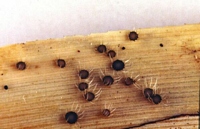
Owner: Peter Johnston | 
Caption: Fig. 9 Hispidula tokerau (A, B = PDD 56335, C-E = PDD 55317). A, B, macroscopic
appearance of apothecia;C, apothecium in vertical section; D, detail of outer ex | 
Caption: Fig. 10 Hispidula tokerau (PDD 56335). A, asci; B, tips of paraphyses; C, released
ascospores; D, distribution of collections examined. Scale bar: A-C = 20 µm. |
Article: Johnston, P.R. (2003). Hispidula gen. nov. (Helotiales, Hyaloscyphaceae) in Australia and New Zealand. New Zealand Journal of Botany 41(4): 685-697 (http://www.rsnz.org/publish/abstracts.php).
Description: DESCRIPTION: Apothecia developing on lower surface of fallen leaves, typically near base
of leaf, associated with bleaching of the leaf tissue. Apothecia erumpent from beneath
epidermis, arising from small patch of compact, hyaline tissue comprising tangled 2-3-µm-diam. hyphae
with thin, hyaline walls, embedded in hyaline gelatinous matrix. Apothecia
0.5-0.8 mm diam., sessile, disc pale yellowish,receptacle pale yellowish to dark brown, with a
row of massive, tapering, pale, whitish appendages up to 0.8 mm long. Ectal excipulum two-layered;
outer layer 50 µm thick, of textura angularis with elements oriented at high angle to
receptacle surface, comprising angular, 7.5-12.5-µm-diam. cells with walls slightly thickened,
hyaline, slightly gelatinous,outermost cells encrusted with bright yellow-brown material (this
dissolving in lactic acid); inner layer up to 20 µm thick near margin of disc, 5-10 µm at sides
of receptacle, comprising long cylindric, 3-µm diam.cells with walls thick, hyaline,
gelatinous. Medullary excipulum poorly developed, barely differentiated from the basal tissue
within the host leaf. Excipular hairs arising from outer layer of ectal excipulum; up to 800 x 3
µm, cylindric with rounded apex, walls thick, smooth, hyaline, dextrinoid in Melzer's reagent
(under interference contrast microscopy with amyloid appearance at some angles), numerous
hairs aggregated into tight groups, forming triangular, tapering appendages, 40 µm wide at
base. Subhymenium comprising tangled 2-3-µm-diam. hyphae with walls hyaline,
thin, nongelatinous. Paraphyses 1.5 µm diam., apex slightly swollen to 2.5-3 µm diam.,
sometimes branched in the upper 50 µm, extending 5-10 µm beyond asci, embedded in
common gelatinous matrix. Asci 80-105 x 6.5-7.5 µm, cylindric to subclavate, apex more or
less broadly rounded, wall slightly thickened at apex, apical pore nonamyloid, 8-spored, spores
overlapping uniseriate, spores extending 65-70 µm from ascus apex. Ascospores 10.5-14 x
3.5-4.5 µm, elliptic to fusoid, ends more or less acute, slightly flattened one side, not
curved, 0-1-septate, wall hyaline (becoming pale brown after ascospores released).
Notes: ETYMOLOGY: The specific epithet refers to the Maori word for the northern part of New
Zealand, tokerau, reflecting the northern distribution of this species (epithet in the form of a
noun in aposition).
NOTES: H. tokerau is macroscopically very similar to H. pounamu. This second species is
distinguished macroscopically by its brighter hymenium when fresh, its larger asci and
ascospores, and by its ascus wall being thicker at the apex, and the apical pore being amyloid.
H. tokerau is known only from the northern part of the North Island of New Zealand.
|