|
 Lanzia novae-zelandiae Lanzia novae-zelandiae
SynonymsLanzia prasina var. nigripes
Helotium novae-zelandiae
BiostatusPresent in region - Indigenous. Non endemic
Images (click to enlarge)
Caption: FIG. 21. Helotium novae-zelandiae. Habit sketch x 10, details x 660. | 
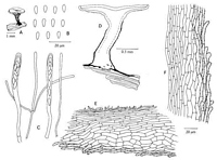
Caption: Figure 61. Lanzia prasinum var. nigripes, holotype. A. Apothecium. B. Ascospores. C.
Asci and paraphyses. D. Vertical section. E. Ectal excipulum on receptacle. F. Ectal
excipulum on stipe. |
Article: Spooner, B.M. (1987). Helotiales of Australasia: Geoglossaceae, Orbiliaceae, Sclerotiniaceae, Hyaloscyphaceae. Bibliotheca Mycologica 116: 711 p.
Description: STROMA substratal, forming irregular, sometimes extensive blackened areas. APOTHECIA
scattered to gregarious, erumpent, stipitate. DISC 1-2 mm diam., concave or plano-concave,
pale yellow when dry, paler and whitish when rehydrated, smooth. RECEPTACLE shallow
cupulate, brown to dark brown when dry, paler and yellowish when rehydrated but with
pigmented fibrils forming conspicuous dark brown vertical streaks, particularly in the lower
part. STIPE central, tapered, concolorous above, dark brown fibrillose, darker downwards,
expanded and dark brown or blackish at the base, 1.5-2.0 mm long. ASCI 8-spored, narrowly
cylindric-clavate, tapered below to a fairly long, narrow base, expanded to a small foot 3-4 µm
diam., apex narrowed, rounded, the pore not staining in Melzer's reagent, (70-)75-81(-85) x 5-6(-7) µm. ASCOSPORES ovate or ovate ellipsoid, sometimes pyriform, broadest above
centre, ends rounded, hyaline, non-septate, uniseriate or partially biseriate. Free spores in one
collection budding subglobose to broadly ellipsoid, hyaline secondary spores from the lower
end or laterally towards the lower end. Ascospores 5.8-8.0 x 2.5-3.0, mean 6.9 (SD 0.7) x 2.7
(SD 0.2) µm. Secondary spores 2.5-3.0 x 2.0-2.5 µm. PARAPHYSES hyaline, filiform,
obtuse, simple or branched in lower part, 1.0-1.5 µm diam., slightly enlarged to 2.0-2.5 µm at
the apex, not exceeding the asci. SUBHYMENIUM ill-defined, composed of hyaline,
interwoven hyphae 1.5-2.0 µm diam. MEDULLARY EXCIPULUM in the stipe composed of
virtually hyaline, compact, vertically orientated, irregularly septate hyphae 2.0-2.5 µm diam.,
with thin or slightly thickened walls; in the receptacle composed of interwoven hyphae 1.5-2.5
µm diam., hyaline or very slightly pigmented, the tissue narrowing towards the margin and
disappearing at the base of the edge of the hymenium. ECTAL EXCIPULUM duplex in the
receptacle. Innermost layer composed of parallel, undulating hyphae similar to those of the
stipe medulla, 80-85 µm thick at the base of the receptacle, narrowing into the margin and
merging with the ectal excipulum to form a layer 20-25 µm thick on the flanks of the
hymenium. Outermost layer 40 µm deep on the stipe, slightly narrower on the receptacle,
composed of broader, shorter celled hyphae forming prismatic cells 10-18 x 6-8 µm, in rows
at a low angle to the surface, narrowing towards the margin and towards the surface, the
outermost 2-3(-5) layers of hyphae being distinctly brown pigmented, paler towards the
margin. Surface hyphae terminating in free, rounded often reflexed tips 2-5 µm diam., the
pigment of the terminal 2-4 cells being irregularly deposited to give a granular appearance.
Hyphal endings broader and more conspicuous on the stipe, disappearing towards the margin.
Habitat: On decorticated wood.
Distribution: Australia, New Zealand.
Notes: The three collections of this taxon from Victoria agree closely in most respects with the type
material from New Zealand. However, in two of them, Beaton 13 and Beaton 95, the
ascospores are budding, though not whilst still within the ascus. This is best observed in
Beaton 13, in which the secondary spores are nearly always budded at or near the distal end of
the spore. They are subglobose or broadly ellipsoid and frequently truncate at the point of
attachment.
Dennis (1961) compared the structure of this taxon with that of Rutstroemia fusco-brunnea
(Patouillard & Gaillard) Le Gal, which has similar but narrower and often slightly curved
ascospores. The latter has been redescribed by Dumont (1981), who has shown the ectal
excipulum to be composed of globose cells and has transferred the name to Moellerodiscus.
Amongst the species referred by Dennis (1964) to Hymenoscyphus series Prasinum, one other
species, H. microspermus (Speg.) Dennis from Argentina, has small ascospores. This was
referred to Rutstroemia by Gamundi (1962). From the description and figures she has
supplied, the species is very similar to the present taxon, having yellowish apothecia with
superficial dark brown fibrils, but less clavate and smaller ascospores 3.2-4.8(-5.6) x (1.3-)1.6-2.4 µm. The dark brown superficial hyphae have granulate walls, and a basal stroma is present.
Clearly the species belongs in the Sclerotiniaceae. The single collection at Kew, cited by
Gamundi, closely matches the type description and has, as she has described, caespitose
apothecia which arise from a common sclerotioid mass. The species probably belongs in
Lanzia, though such development is not characteristic of the genus. It does not occur in L.
prasinum, and it is certain that these taxa are specifically distinct.
Helotium ambiguum Rick, on dead wood from Brazil, was described as having ascospores 7
x 3 µm. Dumont (1981) was unable to locate type material, so the taxonomic position of this
species, and its possible relationship with L. prasinum, remains uncertain. Lanzia ambigua
(Bresadola & Hennings) Carpenter, also from Brazil, is quite distinct on account of its brown
apothecia up to 4 mm diam. and ascospores, 12-15 x 2.5-3.0 µm.
[notes from Lanzia prasinum description]
The small ascospores of L. prasinum are unusual but not diagnostic as a single criterion.
Helotium novae-zealandiae is a very similar taxon which exhibits comparable stromatic
development and cannot be distinguished from L. prasinum on ascospore characters alone.
Dennis (1964) considered it to be a synonym. However, there are structural differences
between these taxa which are reflected in the external appearance of the apothecia. Dried
apothecia of L. prasinum bear an olive-yellow furfuraceous covering which microtome
sections reveal to be due to ectal hyphae which terminate in clavate, free tips which are hyaline
or very faintly pigmented. The disc of this taxon was described by Massee (1901) as
chlorinous and, when rehydrated, now appears pale lemon-yellow. The margin is crenulate
and the receptacle yellow or yellowish brown, becoming lemon-yellow towards the margin,
the stipe is largely concolorous, being dark only at the extreme base. There are three
collections in K, in addition to the type, which are referable to H. novae-zealandiae. These
agree well with the type collection and differ from L. prasinum in having a yellow disc which
is whitish when rehydrated, and in lacking an olive yellow furfuraceous surface. Instead, the
receptacle is yellow, streaked conspicuously with dark brown or blackish fibrils which are
most densely set in the lower part. The stipe is slightly downy and either dark brown to
blackish throughout, or pale only at the extreme apex. The dark brown ectal hyphae of the
stipe terminate in concolorous, cylindrical or only slightly clavate free tips. The striate
appearance of the receptacle is produced by adpressed, cylindrical, 1-2-septate hair-like
hyphae. These collections differ further from L. prasinum in having asci with an apical pore
which does not stain blue in Melzer's reagent. Though Massee (1901) described the asci of L.
prasinum as non-staining, the pore is, in fact, clearly outlined blue in Melzer's reagent.
Lanzia prasinum and H. novae-zealandiae are undoubtedly very closely related, but
available evidence indicates that they can be distinguished. The observable differences seem
unlikely to be the result of variation within a single taxon, but are scarcely sufficient to warrant
recognition of two species. I propose, accordingly, to treat H. novae-zealandiae as a variety
of L. prasinum. Because H. novae-zealandiae has proved not to be restricted to New
Zealand, and as the visible distinction between the taxa is one of colour, I propose a new name
Lanzia prasinum var. nigripes for this taxon.
Article: Dennis, R.W.G. (1961). Some inoperculate Discomycetes from New Zealand. Kew Bulletin 15(2): 293-320.
Notes: This fungus bears considerable resemblance to H. vernalis Dennis but has smaller more
clavate ascospores. The excipulum is formed of rows of large, short, thin-walled cells, up to
12 µ wide, terminating in short adpressed fibrils. Rutstroemia fusco-brunnea (Pat. & Gaill.)
Le Gal is similar in structure but has a very long slender stipe.
|