|
 Hispidula dicksoniae Hispidula dicksoniae
SynonymsHispidula filicum
Hispidula tasmanica
Cyathicula dicksoniae
BiostatusPresent in region - Indigenous. Non endemic
Images (click to enlarge)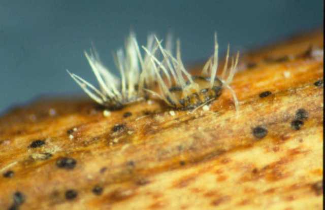
Owner: Landcare Research | 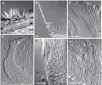
Caption: Fig. 3 Hispidula dicksoniae (PDD 70965). A, macroscopic appearance of apothecia; B,
apothecium in vertical section,one side only; C, detail of side of receptacle, in vertical
s | 
Caption: Fig. 4 Hispidula
dicksoniae (PDD 70965). A, asci; B, tips of paraphyses; C, released ascospores; D,
distribution of New Zealand collections examined. Scale bar |
Article: Johnston, P.R. (2003). Hispidula gen. nov. (Helotiales, Hyaloscyphaceae) in Australia and New Zealand. New Zealand Journal of Botany 41(4): 685-697 (http://www.rsnz.org/publish/abstracts.php).
Description: DESCRIPTION: Apothecia developing on dead fronds, erumpent from beneath epidermis,
with flap of host tissue pushed to one side at base of apothecium, arising from small patch of
compact, hyaline tissue confined to upper few layers of host cells across base of apothecium.
Apothecia 0.5-1 mm diam., sessile, disc pale yellowish, receptacle pale to dark brown, with a
row of massive, tapering, pale, whitish appendages up to 0.8 mm long. Ectal excipulum
twol ayered; outer layer up to 50 µm thick, of textura angularis to textura prismatica with
elements oriented at high angle to receptacle surface, comprising brick-shaped to angular,
6-10-µm-diam. cells with walls thick, hyaline, encrusted with yellowbrown material on
outermost cells, gelatinous; inner layer up to 50 µm thick near edge of disc, 10-15 µm at sides
of receptacle, comprising long cylindric, 2.5-3-µm-diam. cells with walls thick, hyaline
(outermost cells encrusted with yellow-brown material), gelatinous. Medullary excipulum
comprising 65-µm-deep layer of cylindric, partly tangled cells oriented more or less
perpendicular to host surface,4-6 µm diam., with walls slightly thickened, hyaline, slightly gelatinous. Excipular hairs
arising from outer layer of ectal excipulum, up to 800 x 3 µm, cylindric with rounded apex,
walls thick, smooth, hyaline, dextrinoid in Melzer's reagent (under interference contrast
microscopy with amyloid appearance at some angles), numerous hairs aggregated into tight
groups forming triangular, tapering appendages, 60-80 µm wide at base. Subhymenium
comprising 2-4-µm-diam. hyphae with walls hyaline, thin, nongelatinous. Paraphyses 1.5 µm
diam., apex slightly swollen to 2.5-3 µm diam., unbranched, extending 15-30 µm beyond asci,
embedded in common hyaline gelatinous matrix. Asci 90-115(-130) x 5.5-7 µm, cylindric,
tapering slightly to subtruncate apex, wall thickened at apex, apical pore sometimes amyloid,
8-spored, spores overlapping uniseriate or biseriate, spores extending 65-80 µm from ascus
apex. Ascospores 10-14.5 x 3-3.5 µm, elliptic to fusoid, ends more or less acute, flattened one
side, often slightly curved in side view, 0-1-septate, wall hyaline.
Notes: NOTES: H. dicksoniae is macroscopically similar to the other pale-coloured Hispidula spp.,
but can be distinguished by its narrower ascospores, and its substrate. Beaton & Weste (1977)
placed this fungus in Cyathicula. They noted that they did so with "some hesitation", but that
there was no other satisfactory genus for their fungus. Although some Cyathicula species have
large, tooth-like appendages around the margin of the disc, these are simply extensions of
the excipular tissue, and have the same anatomical structureas the highly gelatinised
excipulum of this genus. The apothecial appendages of H. dicksoniae and the other Hispidula
species comprise anatomically distinct hairs. Carpenter (1981) excluded H.dicksoniae from
Cyathicula, compared its excipular structure with Hymenoscyphus, but otherwise made no
attempt to recombine this species. An additional Tasmanian collection (Mt Field National
Park, Lyrebird Nature Walk, on Dicksonia antarctica dead fronds, P. R. Johnston Tas 129
& A. K. Mills, 26 May 1988, HO, PDD 54963) from Dicksonia matches H. dicksoniae
macroscopically, but has slightly wider asci (7-8.5 µm) and ascospores (4-4.5 µm). It could
represent a distinct species, but until more material is available from Australia to
enable assessment of variation between populations of these fungi, its identity remains
doubtful
|