|
 Rosellinia communis Rosellinia communis
BiostatusPresent in region - Indigenous. Endemic
Images (click to enlarge)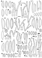
Caption: Fig. 12 A-D, Rosellinia novae-zelandiae, PDD 43205: A, Ascospores, last one immature; B,
Ascus apical ring; C, Conidiophores and conidia on the host (PDD 16422); D, Conidiophores
and conidia in culture (PDD 42074); E-H< | 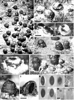
Caption: Fig. 11 Rosellinia communis. A-H, Stromata, B-D, F, showing subiculum, E, concentric rings
on surface; I, Vertical section of stroma, outer shell stroma, inner perithecium; K, A | 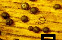
Caption: overhead view of ascostromata in situ on dead decorticated wood; note the dark circular scar where a flat-based superficial ascostroma once rested. Scale bar = 800 µm. | 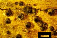
Caption: overhead view of ascostromata in situ on dead decorticated wood; note masses of dark ascospores in some ostiolar areas, discharged as the ascostromata dried out. Scale bar = 800 µm. | 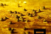
Caption: side view of ascostromata in situ on dead decorticated wood; note their conical shape and concentric rings. Scale bar = 1250 µm. | 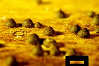
Caption: side view of ascostromata in situ on dead decorticated wood; note their conical shape and concentric rings (closeup from field of photo #3). Scale bar = 625 µm. | 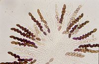
Caption: asci, ascospores and paraphyses; note the longitudinal germ slits on the ascospores. (from SMF mount). | 
Caption: Rosellinia communis: overhead view of ascostromata in situ on dead decorticated wood; note the dark circular scar where a flat-based superficial ascostroma once rested. * = 800 µm. (projection slide #5).
Owner: D. Mahoney | 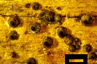
Caption: Rosellinia communis: overhead view of ascostromata in situ on dead decorticated wood; note masses of dark ascospores in some ostiolar areas, discharged as the ascostromata dried out. * = 800 µm. (projection slide #11).
Owner: D. Mahoney | 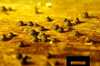
Caption: Rosellinia communis: side view of ascostromata in situ on dead decorticated wood; note their conical shape and concentric rings. * = 1250 µm. (projection slide #15).
Owner: D. Mahoney | 
Caption: Rosellinia communis: side view of ascostromata in situ on dead decorticated wood; note their conical shape and concentric rings (closeup from field of photo #3). * = 625 µm. (projection slide #16).
Owner: D. Mahoney | 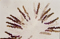
Caption: Rosellinia communis: asci, ascospores and paraphyses; note the longitudinal germ slits on the ascospores. (from SMF mount, projection slide #20).
Owner: D. Mahoney | |
Article: Petrini, L.E. (2003). Rosellinia and related genera in New Zealand. New Zealand Journal of Botany 41(1): 71-138 (http://www.rsnz.org/publish/abstracts.php).
Description: Subiculum evanescent, restricted, approx. to 1 mm in extension, as white, cream patches in
early stages, later light brown, felty, bearing conidiophores, subsequently reduced while
stromata progressively emerge, until absent in old material. Stromata (400)687 ± 119.5(1050)
µm high, (550)804 ± 131(1250) µm wide (n = 134), conical to columnar with bluntly rounded
top, side walls often with concentric rings, wavy (Fig. 11E,H), dark brown, black around the
ostioles, completely black when old, solitary or crowded, touching each other, sometimes 2-3
fused together, when young completely covered by the subiculum, during development
gradually exposed. Ostioles finely papillate to pointed or not pronounced. Ectostroma 50-75
µm thick, black. Entostroma light brown, confined to the base. Perithecia detached and
collapsed in mature material. Ascus apical rings (1.9)2.7 ± 0.5(3.8) µm high, upper width 3.3-
4.8 µm, lower width 2-2.8 µm (n = 63), J+, pale blue. Ascospores (13.4)17.3 ± 1.3(21.6) µm
long, (6.7)8.9 ± 0.7(11.5) µm wide (n = 710), inequilaterally ellipsoidal, dark brown, with
straight germ slit, extending almost over the whole spore length. Conidia 3-4 x 2.5-3 µm.
Cultures on OA after 13 days at 20°C under diffused daylight 0.7-1 cm diam., white to pale
pink, sterile, after 30 days 2.5-3 cm, flat, densely cottony, white when sterile, grey from
conidial production, reverse white. Conidiophores 100-200 µm long, 3- 4 µm wide, forming a
continuous layer over the colony surface, mononematous, macronematous, loosely and
irregularly branched, smooth, pale olivaceous. Conidiogenous cells 19-60 x 2.5-3 µm when
terminal (n = 21), terminal and intercalary alsobearing terminal and intercalary conidiogenous
loci, geniculate with a circular refractive frill at each point of conidial dehiscence. Conidia
3-4(5) x (2)2.5- 3 µm (n = 44), ovoid to subglobose with a flat, c. 1 µm wide basal frill,
refractive. On CMD after 29 days at 20°C under 12 h dark and 12 h UV and fluorescent light
1.5 cm in diam., pale orange, transparent, aerial hyphae short. Conidiophores 80- 160 µm
high, 1.5-2 µm wide, freely branched, bearing a head of conidia at the tip of each branch,
subhyaline to pale tan. Conidiogenous cells 30-55 x 2-3 µm (n = 9), terminal, sometimes
intercalary, geniculate with a circular refractive frill at each point of conidial dehiscence.
Conidia 3-4(5) x 2-3 µm (n = 44), subglobose to ovate with protuberant, 1 µm wide flat basal
abscission scar bearing a minute frill, smooth, subhyaline. On PDA restricted, white, felty,
forming concentric rings, with large grey areas bearing conidiophores.
ANAMORPH: Geniculosporium.
Habitat: HOSTS: Beilschmiedia tawa, Brachyglottis repanda, Freycinetia baueriana subsp. banksii,
Hedycarya arborea, Macropiper excelsum, Melicytus ramiflorus, Neopanax arboreum,
Nothofagus solandri, Populus sp., Rhopalostylis sapida, Schefflera digitata, Sophora
microphylla.
MATRIX: Corticated or decorticated, heavily decomposed wood.
Notes: ETYMOLOGY: communis (common), referring to the frequent occurrence of this species.
NOTES: Rosellinia communis is characterised by itsconical to columnar, black stromata
covered by a whitish cream subiculum when young. The side walls regularly show concentric
rings, thus giving their surface a wavy appearance. Rosellinia communis can be distinguished
easily from R. johnstonii and R. mammoidea by its larger, differently shaped stromata and
ascospore size. Many specimens of R. communis were assigned to R. mammoidea, as the spore
size erroneously published for the latter by Cooke (1879) corresponds to that of R. communis
ascospores. Cooke (1879) gave 16-18 x 8 µm for the Travers collection (the type of R.
mammoidea), whereas the spores of this specimen actually measure 11-14 x 7-8 µm (see R.
mammoidea below).
The closest species is R. picta (Berk.) Cooke described from Sri Lanka. The type material has
regular, conical to semiglobose stromata lacking wavy side walls and ascospores with pinched
ends. The stroma and ascospore size, however, do not differ among the two species as
revealed by analysis of variance and discriminant analysis, respectively (results not shown).
The type material of R. griseo-cincta Starbäck, R. indica Thind, and R. rickii Bres. show
roughly the same shape for stromata and ascospores; the stromata, however, are larger and
lack the wavy surface and the ascospores are smaller (L. E. Petrinu unpubl. data). Rosellinia
communis differs from R. subiculata by stroma shape, size, and subiculum colour as well as
much larger ascospores (Petrini 1993).
|