|
 Rosellinia novae-zelandiae Rosellinia novae-zelandiae
BiostatusPresent in region - Exotic
Images (click to enlarge)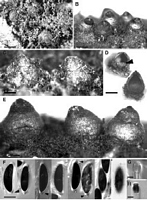
Caption: Fig. 22 Rosellinia novae-zelandiae. A-C, E, Stromata; D, Vertical section of stromata, arrow
points to collapsed perithecium; F, Ascospores, the two to the righ | 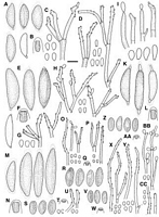
Caption: Fig. 12 A-D, Rosellinia novae-zelandiae, PDD 43205: A, Ascospores, last one immature; B,
Ascus apical ring; C, Conidiophores and conidia on the host (PDD 16422); D, Conidiophores
and conidia in culture (PDD 42074); E-H< | 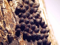
Owner: P. Johnston | 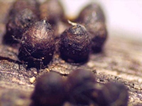
Owner: P. Johnston | |
Article: Petrini, L.E. (2003). Rosellinia and related genera in New Zealand. New Zealand Journal of Botany 41(1): 71-138 (http://www.rsnz.org/publish/abstracts.php).
Description: Subiculum persistent, purplish brown, coarse, wiry, woolly to felty, appressed, when young
producing conidiophores in a grey-brown layer, subiculum reduced in old material. Stromata
(675)899 ± 137(1300) µm high, (625)832 ± 116(1175) µm wide (n = 80), conical to
pear-shaped to ampulliform, often with a short cylindrical base, sometimes with wrinkles on
the surface or faint cracks at the base, dark brown, black around the ostioles, solitary but
densely aggregated forming a compact layer; when young completely embedded in the
subiculum, later gradually exposed and subiculum reduced. Ostioles coarsely papillate, often
poorly differentiated and forming a cone-like top. Ectostroma 50-100 µm thick, black.
Entostroma c. 75 µm thick, cream to black, confined to stroma base. Perithecia detached and
collapsed in mature material, often remaining as a central peg. Ascus apical rings (5.7)7.5 ±
1.2(10.5) µm high, upper width 4-5.7 µm, lower width 4.3- 6.7 µm (n = 63), J+, dark blue.
Ascospores (20)24.3 ± 1.7(30) µm long, (5.7)8.5 ± 0.6(11.5) µm wide (n = 450),
inequilaterally ellipsoidal, brown, with straight germ slit running over the whole spore length,
with a basal, 1.5-2 1.5-2 µm large, cellular appendage, the whole spore completely surrounded
by a slimy sheath, 2 µm thick at extremities, 1 µm thick at the sides. Conidia 6.5-8.5 x 4-5
µm.
Culture on OA after 24 days at 20°C under 12 h dark and 12 h UV and fluorescent light
covering plate (9 cm diam.), mottled with black, effused, stromatic and white, flat, dense
hyphal splotches, cottony, cinerescent hyphae at the contact line between two inocula.
Conidiophores in restricted areas, variable in length, without central axis, repeatedly and
irregularly branched, widely spread, ultimate cells conidiogenous, smooth, subhyaline to pale
tan towards base. Conidiogenous cells 16-43 x 3-3.5 µm (n = 13), disposed in 2s or 3s in a
roughly verticillate fashion, mostly geniculate over c. 10 µm length of the tips, or less
frequent, with swollen tips bearing conidiogenous scars, smooth, with conspicuous, circular
refractive frill at each point of conidial dehiscence. Conidia (4)7-10 x 3- 4 µm (n = 22),
oblong to elliptic with a basal frill, smooth, pale tan.
ANAMORPH: Geniculosporium.
Habitat: HOSTS: Actinidia chinensis, Albizzia lophantha, Beilschmiedia tawa, Euonymus japonicus,
Pittosporum crassifolium, Populus sp., Prunus persica, ?Pseudopanax sp., Rosa sp.
cultivated, Salix caprea, Salix spp., Solanum auriculatum.
MATRIX: Corticated and decorticated wood, twigs, detached bark.
Notes: ETYMOLOGY: Referring to the geographical origin, New Zealand.
NOTES: The most striking feature of R. novae-zelandiae is the pear-shaped, conical stroma
with coarse ostioles that are often poorly differentiated. Cultures of R. novae-zelandiae
resemble those of R. aquila and R. corticium as all develop black discolorations of the colony.
The conidial sizes of the anamorph of R. novae-zelandiae and R. corticium are similar.
Conidia of R. aquila are smaller than those of the other two species (Petrini 1993).
Many specimens previously identified as R. aquila are R. novae-zelandiae. The latter has
larger ascospores with one cellular appendage completely surrounded by a slimy sheath.
Ascospores of R. aquila have two cellular appendages surrounded by slimy caps (Petrini
1993). The ascospores of R. novae-zelandiae are similar to those of R. caudata Petch, R.
corticium (Schwein. : Fr.) Sacc., R. immersa Petch, and R. merrilli Syd., but the stromatal
shape of these species is subglobose to semiglobose and they have papillate to pointed
ostioles. Mature spores of the type material of R. caudata have no cellular appendage and
have a rather thick epispore; those of R. immersa are larger, as illustrated by the 65%
confidence ellipses (Fig. 9D). R. merrillii has larger stromata, larger ascus apical rings, and
larger ascospores (Petrini 1993; L. E. Petrini unpubl. data).
|