|
 Hypohelion scirpinum Hypohelion scirpinum
SynonymsHypoderma scirpinum
BiostatusAbsent from region
Images (click to enlarge)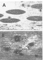
Caption: Fig. 1. Macroscopic appearance of ascomata (x15). A, Hypohelion scirpinum (Rehm, Asc. No. 1953 - NY).
B, H. parvum (PDD 48879) | 
Caption: Fig. 2. Hypohelion scirpinum. A-D, Ascomata in vertical section at different stages of maturity.
(A-D, Fungi Dakotenses No. 142 - NY). | 
Caption: Fig. 3. Hypohelion scirpinum. A, Asci. B, Apex of asci and paraphyses. C, Released ascospores.
D, Conidioma in vertical section. E, COnidiogenous cells and conidia. (A-C, Fungi Dakotenses No. 142 - NY. D-E,
coll. Jackson, 4.x.1933 - NY ex TRTC |
Article: Johnston, P.R. (1990). Hypohelion gen. nov. (Rhytismataceae). Mycotaxon 39: 219-227.
Description: Ascomata developing in slightly paler areas of the leaves, sometimes surrounded by narrow,
black zone lines. Associated with anamorph conidiomata.
In surface view ascomata 0.8-3(-7) x 0.5-0.8 mm, elliptic to broad-elliptic in outline, with
rounded ends. In unopened ascomata the wall is uniformly black, usually developing a fold-like ridge along the long axis of the ascomata. Ascomata opening by an elongate split which
usually forms more or less along the fold-like ridge. No differentiated zone visible along the
edge of the opening split. Parts of the upper wall adjacent to the opening split often break off
and fall away, leaving the hymenium partially exposed.
Ascomata develop between the host cuticle and epidermis, although when ascomata develop
over stomata in the host leaf, hyphae invade and break down a few host cells immediately
below the stomata. In vertical section, at an early stage of development, the ascomatal initial
comprises 2-3 layers of hyaline plectenchyma, covered with a single layer of brown, thick-walled, angular cells, which comprises the first stage in the development of the upper wall
(Fig. 2A). As the ascomata develop the upper wall thickens to 20-30 µm, comprising thick-walled, brown, angular cells, and a cavity develops within the hyaline plectenchyma, with
paraphyses growing upward from the base of this cavity (Fig. 2B). From this stage the upper
wall does not become thicker, but it darkens and becomes distorted and folded by the pressure
exerted from the developing hymenium. The upper wall shows no internal differentiation,
comprising uniformly thickened and darkened, angular cells, but shows a distinct upward fold
near the centre of the ascomata (Fig. 2C). The upper wall splits open in the region of this fold
(Fig. 2D). No layer of differentiated cells is present along the edge of the opening split. A
darkened lower wall does not develop, the hyaline tissue below the hymenium resting directly
on the more or less intact epidermal cells of the host.
Paraphyses 1.5 µm diam., swelling more or less abruptly to 4-6 µm diam. at the clavate to
knob-like apex. Asci 130-160 x 15-18 µm, clavate, tapering gradually to rounded apex, wall
at apex sometimes slightly thickened with indistinct central pore, 8 spored, spores extending
two-thirds down the ascus, maturation sequential. Ascospores 40-75 x 4.5-6.5 µm, tapering
slightly to both ends, 0-1 septate, surrounded by narrow gelatinous sheath.
Conidiomata 0.2-0.3 mm diam., round in outline, dark brown to black, pustulate,
subcuticular. In vertical section lenticular in shape, upper wall 3-5 µm thick, comprising dark
brown material without obvious cellular structure. Darkened lower wall absent, the
conidiogenous layer developing on 2-3 rows of thin-walled, hyahne, angular cells.
Conidiogenous cells 9-15 x 2-3 µm, cylindric, solitary, forming a palisade-like layer, with
sympodial proliferation, often with more than one developing conidium held at the apex.
Conidia 3-4.5 x 1 µm, oblong-elliptic with rounded ends, or short-clavate with a broadly
rounded apex and tapering to a truncate base, hyahne, 0 septate.
Habitat: Dead leaves of Scirpus.
Distribution: Northern Europe and northern North America (Powell 1974).
|