|
 Pirottaea nidulans Pirottaea nidulans
SynonymsPirottaea falcata
Chaetoscypha nidulans
BiostatusPresent in region - Indigenous. Endemic
Images (click to enlarge)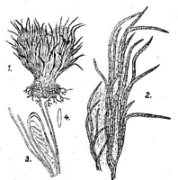
Caption: Fig. 2. Chaetoscypha nidulans Syd.
1. Seitenansicht eines Apotheziums. Vergr. 70:1.
2. Teil der Wandung desselben. Vergr. 260:1.
3. Schlauch mit Paraphysen. Vergr.
260:1.
4. Spore. Vergr. 260:1. | 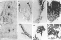
Caption: Fig.1 Pirottaea falcata (A-F, H, PDD 62688; G, PDD 49112): A, apothecia; B, apothecium
dislodged and lying on leaf tomentum, showing short stipe; C, apothecium in vertical section;
D, margin of | 
Caption: Fig. 2 Pirottaea falcata (A-C, PDD 62647; D, PDD 68331): A, apex of paraphyses; B, asci;
C, ascospores; D, falcate setae from margin of disc, in surface view; E, 
Caption: Fig. 1 Pirottaea falcata as illustrated by Johnston (1998). (Original caption: Fig. 1 Pirottaea
falcata (A-F, H, PDD62688; G, PDD 49112.) A, apothecia; B, apothecium dislodged and
lyin | | 
Caption: Fig. 2 Chaetoscypha nidulans as illustrated by Sydow(1924). (Original caption: Fig. 2.
Chaetoscypha nidulansSyd. 1. Seitenansicht eines Apotheziums. Vergr. 70:1.; 2.Teil der
Wandung desselben. Vergr. 260:1.; 3. Schlauc |
Article: Johnston, P.R. (1998). Four new Pirottaea species from New Zealand. New Zealand Journal of Botany 36(4): 645-652 (http://www.rsnz.org/publish/abstracts.php).
Description: Apothecia developing within leaf tomentum on undersides of living leaves of Olearia species (Asteraceae). Apothecia 0.1-0.15 mm diam., 0.2 mm high, black with numerous stiff, black setae, urn-shaped, tapering suddenly to a short, stipe-like base. Setae near top of apothecium extending outwards across top of leaf tomentum.
Ectal excipulum 2-layered. Inner layer 6-10 µm wide, in vertical section comprising 2 or 3 rows of brick-shaped cells 4.5-6 µm diam., with thin, brown walls. Outer layer patchy in development, comprising a single layer of cylindric cells 2.5-3 µm diam., with pale brown, irregularly encrusted walls, and with setae arising from these cells. Setae mostly near top of apothecium, 60-80 x 5-6.5 µm, often distinctively sickle-shaped, with very dark, thick walls, probably 0-septate. Seta-like or hair-like elements at sides and near base of apothecium, 12-20 x 2-3 µm, irregularly cylindric, with brown, slightly thickened walls, usually 2 or 3-septate. Medullary excipulum poorly developed, comprising 2 or 3 rows of hyaline, narrow-cylindric cells.
Paraphyses 2-2.5 µm diam., undifferentiated to slightly swollen near apex, about same length as the asci. Asci 55-70 x 6.5-7.5 µm, cylindric, tapering to slightly truncate apex, wall thickened at apex, non-amyloid, 8-spored. Ascospores 6.5-8 x 3-4 µm, broadly rounded at both ends, subclavate, slightly wider in upper half, hyaline, 0-septate.
Cultures approximately 50 mm diam. after 4 weeks. On OA with low, dense, felted,
pale pinkish-grey mycelium; yellowish in reverse. On MEA with dense, cottony, white to
pale grey mycelium; grey brown in reverse. Cultures remained sterile.
Notes: ETYMOLOGY: falcata, refers to the sickle-shaped setae.
NOTES: Together with the unusual host substrate, ascospore size and shape differentiate C.
falcata from the species treated by Nannfeldt (1985). Although most of these were also
found on Asteraceae, all developed on dead leaf and stem tissue rather than the tomentum of
living leaves. The asci of P. falcata appear to mature and discharge their spores while the
leaves of the host plant are still alive, since all apothecia examined from fallen leaves were
over-mature.
Several other species of Leotiales are associated with the leaf tomentum of Asteraceae
in New Zealand. Johnston (1989) described three Crocicreas species and recorded C.
epitephrum (Berk.) S.E. Carp., first described from Australia, from the leaf tomentum of
various species. Another Australian species, Lachnum willisii (G.W. Beaton) Spooner,
described from the leaf tomentum of Celmisia asteliifolia from Australia, also occurs on the
tomentum of living leaves of Celmisia in New Zealand (MID CANTERBURY: Craigieburn
Forest Park, near skifield, on Celmisia discolor, P.R. Johnston & E.H.C. McKenzie, 24 Feb
1988 (PDD 48634); Craigieburn Forest Park, near skifield, on Celmisia angustifolia, P.R. Johnston & E.H.C.
McKenzie, 24 Feb 1988 (PDD 48458). NORTH CANTERBURY: Arthurs Pass National
Park, Nature Trail near Temple Basin, on Celmisia discolor var. intermedia, P.R. Johnston &
E.H.C. McKenzie, 25 Feb 1988 (PDD 48461). WELLINGTON: Tararua Ranges, vic. Dundas
Hut, Pukemoremore summit, on Celmisia sp., P.R. Johnston, 10 Feb 1985 (PDD 49055)), and
there remain many other undescribed leotiaceous species on this substrate in New Zealand
(P.R. Johnston, unpubl. data).
Article: Sydow, H. (1924). Beiträge zur Kenntnis der Pilzflora Neu-Seelands - I. Annales Mycologici 22(3-6): 293-317 Berlin:.
Notes: Die im dichten Blattfilze nistenden schwarzen, punktförmigen Fruchtkörper erwecken zunächst durchaus nicht den Eindruck eines Discomyzeten, ,doch ist deren wahre Natur unter dem Mikroskop sofort zu erkennen. Durch die starren dunkel gefärbten Borsten erinnert der Pilz etwas an Pirottaea. Diese Gattung besitzt jedoch durchweg parenchymatisch gebaute Gehäuse, während die Gehäuse des vorliegenden Pilzes durchaus einen prosenchymatischen Aufbau erkenhen lassen. In Betracht zu ziehen sind auch die Gattungen Dasyscypha und Unguicularia. Beide Gattungen enthalten jedoch fast aussehliesslich hell gefärbte Pilze. Ausserdem sind die Dasyscyphen mit meist septierten, stumpfen Haaren, aber nicht mit so starren, zugespitzten Borsten besetzt, wie solche der neue Pilz aufweist. Unguicularia weist zwar zugespitzte 1-zellige Haare auf, doch sind dieselben hier hyalin und auch wesentlich anders beschaffen. Der kleine völlig oberflächlich wachsende neuseeländische Pilz muss daher als Vertreter einer eigenen Gattung angesehen werden.
Zu beachten ist noch, dass die im oberen Teil völlig frei stehenden Borsten des neuen Pilzes sich nach unten bis fast zum Grund des Gehäuses verlängern und auf diese Weise die äussere Wand der Apotbezien bilden, die demnach langfaserig und dunkel erscheint. Darunter liegt eine hellere, aus kürzeren, faserigen Zellen gebaute Innenschicht.
Article: Johnston, P.R. (2002). Chaetoscypha Syd. reassessed. New Zealand Journal of Botany 40(4): 697-699 (http://www.rsnz.org/publish/abstracts.php).
Notes: Johnston (1998) described Pirottaea falcata from the leaf tomentum of Asteraceae in New
Zealand (from species of both Olearia and Celmisia). The descriptions and illustrations of
Johnston (1998; Fig.1) and Sydow (1924; Fig. 2) clearly represent the same fungus,
characterised by small apothecia witha short, stipe-like base, and sickle-shaped
setae, embedded within the leaf tomentum on the undersides of leaves of Asteraceae. The
ascospore sizes given in Sydow's (1924) description are slightly longer and narrower than
those given by Johnston (1998). This may reflect differences in mounting media used in the
studies, or could reflect differences in the level of maturity of the material examined. Johnston
(1988) saw ascospores only within the asci, possibly indicating that the material examined was
slightly immature. The type specimen of Chaetoscypha nidulans was not able to be examined.
Although listed in the PDD herbarium catalogue, the type specimen of C.nidulans was noted
as missing from PDD by McKenzie et al. (1992). Duplicates of this specimen are not present in
either of the European herbaria with major Sydow holdings, S and W.Sydow (1924)
considered that C. nidulans could not be placed in the genus Pirottaea Sacc. because of what
he described as a prosenchymatous excipulum, in contrast to the parenchymatous excipulum
that he considered characteristic ofPirottaea. The illustrations of Johnston (1998; Fig.1) show
that the excipular structure in fact appears to match Pirottaea well, although the excipulum
is extremely reduced (in places the ectal excipular layer being a single cell wide), and there is a
poorly developed outer layer comprising meandering hyphae across the surface of the
receptacle. The dark,thick-walled setae are quite unlike the hair-like elements characteristic of
Hyaloscyphaceae. Chaetoscypha was not treated by Nannfeldt (1932,1985) in his detailed
treatments of Pirottaea and related genera.
Sydow (1924) described the genus Chaetoscypha, with the New Zealand fungus C. nidulans
Syd. as the type species. No further species have been added to the genus, and its position
remains uncertain. Kirk et al. (2001) considered that it may be a synonym ofLachnum Retz.,
possibly following Sydow's suggestion that this species belonged in his higher taxonomic
group "Dasyscyphearum", a group similar in concept to the modern
Hyaloscyphaceae. Chaetoscypha nidulans was described from a single collection made from
fallen leaves of the alpine tree daisyOlearia colensoi. C. nidulans develops within the
tomentum on the lower leaf surface, a feature well developed on O. colensoi and many other
New Zealand alpine Asteraceae.
TAXONOMIC TREATMENTPirottaea nidulans (Syd.) P.R.Johnst., comb. nov.ç
Chaetoscypha nidulans Syd., Annales Mycologici 22, 305 (1924).= Pirottaea falcata
P.R.Johnst., New Zealand Journal of Botany 36, 645 (1998).
As Chaetoscypha nidulans is the type (and only) species of Chaetoscypha Syd. 1924, this
genus is now a synonym of Pirottaea Sacc. 1878.
Article: Gadgil, P.D. (in association with Dick, M.A.; Hood, I.A.; Pennycook, S.R.) (2005). Fungi on trees and shrubs in New Zealand. Fungi of New Zealand. Ngā Harore o Aotearoa 4: xi + 437 p. Hong Kong: Fungal Diversity Press.
Description: Type: Foliicolous Fungi; Description: Ascomata apothecial, discrete, superficial, urn-shaped, tapering abruptly to a short, stipe-like base, black, 0.1–0.15 mm in diameter and 0.2 mm high, with numerous stiff, black, sickle-shaped setae; developing within tomentum on the undersides of leaves. Asci cylindrical, 55–70 × 6–8 μm. Ascospores subclavate, 0-septate, 6–8 × 3–4 μm, smooth, hyaline.
Distribution: Distribution: Wellington, Buller, Marlborough Sounds, Chatham Islands.; 1st Record: Sydow (1924: as Chaetoscypha nidulans).
|