|
 Moellerodiscus coprosmae Moellerodiscus coprosmae
BiostatusPresent in region - Indigenous. Endemic
Images (click to enlarge)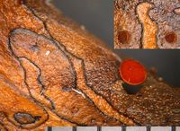
Owner: J.A. Cooper | 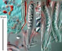
Caption: excipulum, asci, paraphyses and ascospores - in KOH
Owner: J.A. Cooper | 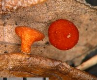
Owner: J.A. Cooper | 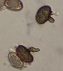
Caption: spores turn brown when germinating on agar
Owner: P.R. Johnston | 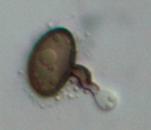
Caption: spores turn brown when germinating on agar
Owner: P.R. Johnston | 
Caption: Fig. 1 Moellerodiscus coprosmae(A-B, PDD
55582; C, PDD 63159; D, PDD 70133; E, PDD 70061; F, PDD 70127). A,
macroscopic appearance with several apothecia in a single, large, irregularly shaped stromatic
area; | 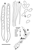
Caption: Fig. 2 Moellerodiscus coprosmae (PDD 62674). A, asci containing ascospores; B, apex of
paraphyses; C, released ascospores; D, distribution of collections examin | 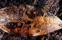
Caption: Dark red discs in stromatic patches on fallen leaves.
Owner: Herb PDD | |
Article: Johnston, P.R. (2002). Three new species of Moellerodiscus (Helotiales, Rutstroemiaceae) from New Zealand. New Zealand Journal of Botany 40(1): 105-115 (http://www.rsnz.org/publish/abstracts.php).
Description: DESCRIPTION: Apothecia developing on upper and lower surface and on petioles of fallen
leaves, on leaf blade associated with irregularly shaped stromatic areas (often with several
areas on a single leaf) about 5-15 mm across, leaf surface in these areas pale grey, margin
marked by narrow, black zone line; these areas contain scattered, round, dark spots 0.1-0.2
mm diam. from which the apothecia arise; infected petioles completely blackened.
Apothecia erumpent from beneath epidermis, arising from basal stroma about 100 µm diam.,
comprising mass of tightly interwoven hyphae 3-7 µm diam. with walls brown to dark brown,
irregularly thickened, refractive, sometimes with scattered groups of crystals 20-40 µm diam.
around edges of stroma; hyphae within host tissue sparse, with walls thickened, refractive, pale
brown to hyaline, intermixed with the still partially intact cells of the host leaf. Apothecia
1.5-5 mm diam., cupulate, short stipitate to stipitate; disc plane, very dark red when fresh,
black when dry; receptacle finely roughened, concolorous with disc when fresh, when dry very
dark red to black with fine, bright rusty brown flecks on the surface; stipe cylindric,
concolorous with receptacle. Ascomata release deep purplish red ("very deep Red",
Kelly 1965) pigment in KOH. Ectal excipulum 30 µm thick on receptacle, textura globosa to
textura angularis with elements oriented more or less perpendicular to receptacle surface,
comprising short cylindric, angular or globose, 5-12 µm diam. cells with walls thick, hyaline,
refractive, outermost cells 6-10 µm diam., more or less free, containing deep purplish red
pigments. Medullary excipulum textura intricata of hyphae 5-6 µm diam. with walls
thin, encrusted, pale brown, nongelatinous (toward base of stipe cells with walls slightly
thickened, refractive,darker). Subhymenium a dense textura intricata comprising hyphae with
walls pale brown, thin, nongelatinous. Paraphyses 2 µm diam., slightly swollen to rounded
apex, 2.5-4.5 µm diam.,unbranched, containing bright orange-brown pigments in water,
changing to deep purplish red in KOH, about same length as asci. Asci (70-)80-95(-100) x
(7.5-)8.5-9.5(-10.5) µm, cylindric to subclavate, tapering slightly to subtruncate apex, wall
thickened at apex, apical pore amyloid with reaction extending through wall, more intense
to inside and outside of wall, 8-spored, spores overlapping uniseriate, extending 55-65 µm
from ascus apex. Ascospores (7.5-)9-10(-11) x (4-)4.5-5.5(-6) µm (x- 9.5 x 4.75 µm, n = 48), broad
cylindric ovate, rounded at both ends, slightly curved, flattened one side, often slightly wider in
upper half, 0-septate, wall hyaline, thin, smooth.
Notes: ETYMOLOGY: Refers to host substrate.
NOTES: Moellerodiscus coprosmae is macroscopically similar to M. griseliniae (see
Discussion and Table 1).In some collections of M. coprosmae, objects that have the
appearance of released ascospores, but with brown walls, have been seen on top of the
hymenium and on the surface of the receptacle. This feature could be used to infer a
relationship withLambertella (cf. Hosoya & Otani 1997); however, many apparently unrelated
sclerotiniaceous discomycetes have ascospores that turn brown following release (see, for
example, Johnston &Gamundí 2000), and this does not appear to be a "good" generic
character. The type species ofLambertella, L. corni-maris Höhn., has an excipular structure
quite unlike Moellerodiscus, the ectal excipulum comprising long-celled elements oriented at a
low angle to the receptacle surface.
|