|
 Terriera nematoidea Terriera nematoidea
SynonymsLophodermium nematoideum
BiostatusPresent in region - Indigenous. Endemic
Images (click to enlarge)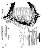
Caption: Fig. 5 Lophodermium nematoideum. A, ascoma in vertical section B, asci. C, apices of asci
and paraphyses. D, released ascospores. (A-E, PDD 54132.) | 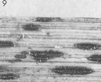
Caption: Fig.9 Macroscopic appearance of ascomata (xl5). Lophodermium nematoideum (PDD54132). | 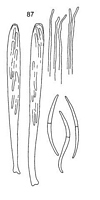
Caption: Fig. 87. Terriera nematoidea (PDD 54132). Asci,
apex of paraphyses, and ascospores. (bar = 20 µm). |
Article: Johnston, P.R. (1991). Rhytismataceae in New Zealand. 4. Pureke zelandicum gen. and sp. nov. plus additional species in Hypoderma, Lophodermium, and Propolis. New Zealand Journal of Botany 29: 395-404 (http://www.rsnz.org/publish/abstracts.php).
Description: Ascomata and conidiomata developing in discrete groups in undifferentiated areas of the leaf,
not associated with zone lines. Ascomata 0.7-1.7 x 0.2-0.3 mm, elliptic to sublinear in outline,
ends rounded. Unopened ascomata black-walled, with paler areas toward both ends of
ascomata, and sometimes with indistinct paler zone along the future line of opening. Opened
ascomata black-walled, with black, flattened zone along both sides of the single, longitudinal
opening.
Ascomata initially subepidermal, epidermal cells becoming filled with fungal tissue as
ascomata mature. Scattered fibre cells from the host leaf become trapped within the walls of
the developing ascomata. In vertical section ascomata initially comprising rows of vertically
oriented, hyaline cells, with the lowermost cells becoming dark brown and thick-walled.
Paraphyses develop within a central cavity and, as they elongate, the lower wall becomes
darker, and the epidermal cells become filled with angular, thick-walled, dark brown fungal
cells. In opened ascomata both the upper and lower walls are almost entirely composed of
very dark tissue with no obvious cellular structure, intermixed with fibre cells of the host leaf.
A few pale, thin-walled, cylindric cells remain at the sides of the hymenium.
Paraphyses 1-1.5 µm diam., undifferentiated at apex, not extending beyond asci. Asci 70-80
x 5-6.5 µm, cylindric to subclavate, tapering slightly to broad, truncate apex, wall unthickened
at apex, sometimes with small, central pore, development sequential, 8-spored, spores
confined to the upper 40-50 µm of the ascus. Ascospores 30-35 x 1.5 µm, tapering toward
both ends, curved or coiling on release, 1-septate, with no obvious gelatinous sheath.
Conidiomata 0.3 mm diam, round in outline, pale brown with darker line around outside edge.
In vertical section lenticular in shape, both upper and lower walls 5-10 µm thick, of 1-3 layers
of brown to dark brown, slightly thick-walled, angular cells, the cells of the lower wall darker
than those of the upper. Both upper and lower walls lined with 1-2 rows of hyaline, thin-walled cells, on which the solitary conidiogenous cells are formed. Conidiogenous cells 8-12 x
1.5-2.5 µm, cylindric or tapering to apex, proliferating sympodially, often with two developing
conidia held at apex. Conidia 3-4 x 1 µm, oblong-elliptic with rounded ends, 0-septate,
hyaline.
Habitat: dead leaves of Gahnia sp.
Notes: ETYMOLOGY: nematoideum = nematode-like, refers to shape of released ascospores.
NOTES: Macroscopically L. nematoideum is typical of the group of species with the pattern
of ascomatal development matching L. minus (Tehon) Johnston (Johnston 1989a (as L.
multimatricum Johnston), 1989b). It is easily distinguished microscopically from the other
species in this group by ascospore shape and size, but is indistinguishable macroscopically
from L. minus and L. breve (Berkeley) de Notaris, both also found on Gahnia.
Article: Johnston, P.R. (2001). Monograph of the monocotyledon-inhabiting species of Lophodermium. Mycological Papers 176: 1-239.
Description: Infected areas on dead leaves, slightly paler than surrounding host tissue, associated with
partial zone lines, containing ascomata and conidiomata. Ascomata 0.7-1-7 x 0.2-0-3 mm,
elliptical to sublinear in outline, unopened ascomata wall black, with pale areas near both ends,
and with faint pale zone along future line of opening, single longitudinal opening slit with
black shelf-like zone along either side. Ascomatal insertion subepidermal, sometimes also
beneath scattered host fibre bundles. Ascomatal structure typical of Terriera, but with
darkened lower wall very well developed. Paraphyses 1-1 5 µm diam., undifferentiated at
apex, embedded in gel, extending c. 20 µm beyond asci. Asci 70-80 x 5-6.5 µm, subclavate,
tapering slightly to truncate apex with slightly thickened wall and small central pore, 8-spored,
well-developed basal stalk with spores confined to upper 40-50 µm. Ascospores 30-35 x 1
µm, tapering slightly to ends, gently curved or sigmoid on release, 1-septate, gelatinous sheath
lacking. Conidiomata 0.3 mm diam., round in outline, wall pale brown with darker line
around outside edge, in vertical section upper and lower walls comprising one to three rows of
brown to pale brown cells and lined with one or two rows of thin-walled, hyaline cells on
which conidiogenous cells are held. Conidiogenous cells 8-12 x 1.5-2.5 µm, solitary, tapering
to apex, proliferation sympodial, often with 2 developing conidia held at apex. Conidia 3-4 x
1 µm, oblong-elliptical with ends rounded, non-septate, hyaline.
Habitat: Gahnia (Cyperaceae).
Distribution: New Zealand.
Notes: Terriera nematoidea is distinguished by its short, broad asci with a truncate apex and
short ascospores. Unusually for a species of Terriera, the epithecium is poorly developed.
|