|
 Terriera asteliae Terriera asteliae
SynonymsLophodermium asteliae
BiostatusPresent in region - Indigenous. Endemic
Images (click to enlarge)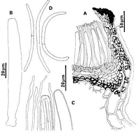
Caption: Fig. 1 Lophodermium asteliae: A, ascocarp margin in vertical section (PDD 43267). B,
ascus (PDD 49088). C, apex of asci and paraphyses (PDD 49088). D, released ascospores
(PDD 49088). | 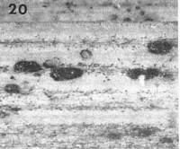
Caption: Fig. 20 Lophodermium asteliae (PDD 48000). Macroscopic appearance of ascocarp (x15). |
Article: Johnston, P.R. (1989). Rhytismataceae in New Zealand. 2. The genus Lophodermium on indigenous plants. New Zealand Journal of Botany 27(2): 243-274 (http://www.rsnz.org/publish/abstracts.php).
Description: Ascocarps and pycnidia developing in pale areas, not associated with zone lines. Ascocarps
0.5-0.8 x 0.3-0.4 mm, elliptic to cylindric in outline, with rounded ends. Immature ascocarps
with dark brown to black walls, some with faint paler zone along future line of opening.
Mature ascocarps with black flattened area along both sides of ascocarp opening. Structures
resembling pycnidia of Rhytismataceae associated with ascocarps, 0.2-0.3 mm diam.,
concolorous except for dark line around outside edge, immersed.
Ascocarps in vertical section initially subepidermal with a layer comprising rows of
hyaline, vertically oriented, cylindric cells surrounding the developing hymenium. As
ascocarps mature the lowermost of these cells become dark brown, and a separate upper wall,
comprising dark brown, thick-walled cells, develops within the host epidermis. A narrow
extension to the upper wall near the ascocarp opening, comprising dark tissue with no obvious
cellular structure covers top of the hymenium.
Paraphyses 1-2 µm diam., undifferentiated at apices, extending 15-20 µm beyond asci.
Asci 75-105 x 8-10.5 µm, cylindric to subsaccate, apex broadly truncate, wall not thickened,
8-spored. Ascospores 45-70 x (1.5-)2-2.5 µm, 0-1 septate, narrow gelatinous sheath, curving
on release. Pycnidia-like structures in vertical section lenticular in shape, upper wall more or
less obsolete, lower wall of 2-3 layers of dark brown, thick-walled cells. Conidia and
conidiogenous cells not seen.
Habitat: Dead leaves of Astelia spp.
Distribution: Northland, Auckland.
Notes: ETYMOLOGY: asteliae; refers to host plant.
NOTES: Collections typical of L. multimatricum have also been found on Astelia. L. asteliae
is easily distinguished by ascus size and shape. See notes under L. multimatricum.
Article: Johnston, P.R. (2001). Monograph of the monocotyledon-inhabiting species of Lophodermium. Mycological Papers 176: 1-239.
Description: Infected areas on dead leaves, paler than surrounding leaf tissue, not associated with zone
lines, containing scattered ascomata, conidiomata not seen. Ascomata 0.5-0.8 x 0.3-0.4 mm,
elliptical to oblong in outline, ends rounded, wall black with poorly-defined paler zone alone,
future line of opening, single, longitudinal opening slit with black flattened zone along either
side. Ascomatal insertion subepidermal. Ascomatal structure typical of Terriera (see p. 37).
Paraphyses 1-2 µm diam., ± undifferentiated at apex, extending 15-20 µm beyond asci. Asci
75-105 x 8-10.5 µm, cylindrical, apex broadly-truncate with undifferentiated wall, 8-spored,
short, broad basal stalk with spores extending almost to base. Ascospores 45-70 x 2-2.5 µm,
tapering slightly to ends and slightly constricted near centre, non- or 1 -septate, gently curved
on release, with no gelatinous sheath.
Distribution: New Zealand.
Notes: Terriera asteliae is characterized within the genus by its broad asci with a broad-truncate apex. Terriera fuegiana, described on Rostkovia from Tierra del Fuego, has similar-sized asci and ascospores. Although the only material of T fuegiana available is in poor
condition, the asci appear to be broadly rounded at the apex, and illustrations by
SPEGAZZINI included in the packet clearly show asci of this shape. Structures resembling
conidiomata of Rhytismataceae were associated with the ascomata, but conidiogenous cells
and conidia were not seen.
|