|
 Dermocybe canaria Dermocybe canaria
SynonymsCortinarius canarius
Cortinarius (Dermocybe) sp.
BiostatusPresent in region - Indigenous. Non endemic
Images (click to enlarge)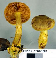
Owner: J.A. Cooper | 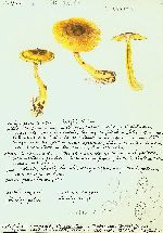
Owner: G.M. Taylor | 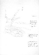
Owner: G.M. Taylor | 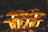
Caption: Peter Johnston | 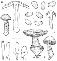
Caption: Dermocybe canaria: 1. carpophores. - 2. spores. - 3. basidia. - 4. cheilocystidia. - 5. carpophores. | 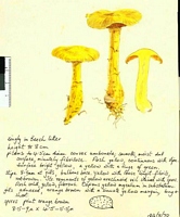
Caption: Watercolour
Owner: G.M. Taylor | 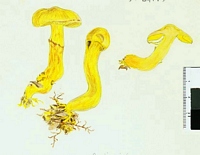
Caption: Watercolour
Owner: G.M. Taylor | 
Owner: J.A. Cooper | 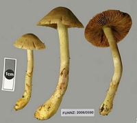
Caption: FUNNZ: 2006/0590, See public note for more information
Owner: FUNNZ | 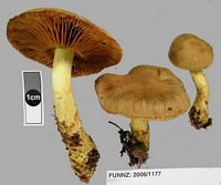
Caption: FUNNZ: 2006/1177, See public note for more information
Owner: FUNNZ | 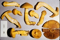
Caption: ZT1901
Owner: E. Horak: © Creative Commons Attribution-Noncommercial 3.0 New Zealand | 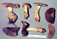
Caption: ZT68-178
Owner: E. Horak: © Creative Commons Attribution-Noncommercial 3.0 New Zealand | 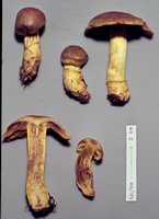
Caption: ZT68-098 , Holotype
Owner: E. Horak: © Creative Commons Attribution-Noncommercial 3.0 New Zealand | 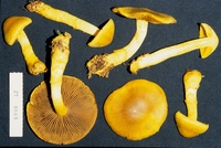
Caption: ZT8669
Owner: E. Horak: © Creative Commons Attribution-Noncommercial 3.0 New Zealand |
Article: Horak, E. (1988) [1987]. New species of Dermocybe (Agaricales) from New Zealand. Sydowia 40: 81-112.
Description: Pileus -60 (-80) mm, hemispherical becoming convex, centre broadly umbonate to campanulate or flat and expanded, margin inrolled; brilliant yellow turning yellow-brown (especially at disk) with age; dry, neither hygrophanous nor striate, minutely to coarsely innate-fibrillose or subsquamulose (in dry condition), in young specimens margin covered by yellow fibrillose veil remnants. - Lamellae crowded, emarginate, sometimes decurrent with short tooth; golden yellow turning mustard yellow then brown-yellow, edges entire, concolorous. - Stipe -80 x -30 mm, fusoid to bulbous with root-like base, rarely cylindrical; yellow, yellow-brown in aged specimens; dry, below yellow cortina with 1-3 persistent, membranaceous, yellow to yellow-brown belts of veil, at first solid becoming hollow, single, rarely cespitose. - Context yellow to orange-yellow. - Odour none. - Taste bitter. - Chemical reactions on pileus: KOH - red to red-brown, NH3-pink to pale orange, HCl - negative.
Spore print (yellow-)brown with rusty shade. - Spores (7-) 7.5-8.5(-9) x 4-5 µm, ovoid, yellow-brown with rusty tinge, minutely asperulate at apex, ± smooth in immature spores, membrane thinwalled. - Basidia 20-35 x 6-8 µm, 4-spored. - Cystidia absent, but edge of lamellae often covered with articulate cells, terminal cell clavate to vesiculose (10-35 x 6-10 µm), membrane hyaline, pigment absent. - Pileipellis a cutis composed of cylindrical hyphae (5-10 µm diam.), terminal cells rarely fusoid, membranes thin-walled, not gelatinized, smooth, with red to orange plasmatic pigment dissolving in KOH, rarely also with encrusting and/or intercellular pigment. - Clamp connections present.
Habitat: On soil among litter in Nothofagus-forests (N. solandri var. cliffortioides, N. menziesii and/or N. fusca; often in association with conifers viz. Libocedrus bidwillii, Phyllocladus alpinus, Ph. glaucus, Podocarpus nivalis both in Sphagnum swamps and on dry sites), rarely also under Leptospermum scoparium. - New Zealand.
Notes: In New Zealand Nothofagus-forests D. canaria is a common species met within a remarkably wide ecological range. This Dermocybe is readily identified by the brilliant yellow colours of the often rather robust carpophores with fusoid to bulbous stipes. The most distinctive microscopical feature are ± smooth spores whose roughened apices are covered with very inconspicuous asperulate warts.
KOH turns the yellow pigment on all parts of the carpophore immediately red. Chromatographic pigment analysis showed that physcion and erythroglaucin are the most abundant anthraquinones of D. canaria. In addition another metabolite, 4-aminophyscion (KELLER & STEGLICH, 1987), was isolated representing the first record of a natural anthraquinone with an amino group.
Adding the macroscopical, microscopical and chemical characters D. canaria obviously occupies an isolated taxonomic position within the infrageneric frame of Dermocybe.
|