|
 Hispidula rubra Hispidula rubra
BiostatusPresent in region - Indigenous. Endemic
Images (click to enlarge)
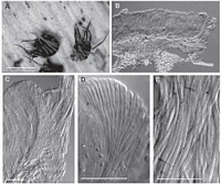
Caption: Fig. 7 Hispidula rubra (A, PDD 70966; B-D, PDD 49023; E, PDD 63173). A, macroscopic
appearance of apothecia; B, apothecium in vertical section; C, detail of side of receptacle i | 
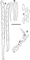
Caption: Fig. 8 Hispidula rubra (PDD
70966). A, ascus; B, tips of paraphyses; C,
released ascospores; D, distribution of collections
examined. Scale bar: A-C = 20 µm. | 
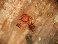
Caption: fruitbody
Owner: J.A. Cooper |
Article: Johnston, P.R. (2003). Hispidula gen. nov. (Helotiales, Hyaloscyphaceae) in Australia and New Zealand. New Zealand Journal of Botany 41(4): 685-697 (http://www.rsnz.org/publish/abstracts.php).
Description: DESCRIPTION: Apothecia developing on both upper and lower surface of fallen leaves,
associated with bleaching of leaf tissue and with reddish staining in the immediate vicinity of
the apothecia. Apothecia erumpent from beneath epidermis, arising from small patch of
compact, hyaline tissue comprising tangled 3-µm-diam. hyphae with walls hyaline, slightly
thickened. Apothecia 0.4-1 mm diam., sessile,disc yellowish when fresh, drying dark
red, receptacle dark red with a row of massive, tapering, dark red to blackish appendages up to
1 mm long. Ascomata release deep wine red pigment in KOH (pigment becoming colourless in
Melzer's reagent). Ectal excipulum two-layered; outer layer up to 65µm thick at sides of
receptacle, of textura angularis to textura prismatica with elements oriented at high angle to
receptacle surface, comprising short cylindric to angular, 6-12-µm-diam. cells with
walls thick, dark red (encrusted with dark red material which dissolves rapidly in KOH),
gelatinous, outermostcells of each element becoming long-cylindric and extending a short
distance along sides of receptacle; inner layer up to 50 µm thick near edge of disc, 10-15 µm
at sides of receptacle, comprising long cylindric to cylindric, 3-µm-diam. cells with walls thick,
hyaline, gelatinous (with red crystal-like inclusions amongst the gel, these dissolving rapidly
in KOH). Medullary excipulum a 60-µm-deep layer of cylindric cells, partly tangled and
oriented more or less perpendicular to host surface, 3-5 µm diam. with walls thick, hyaline,
gelatinous. Excipular hairs arising from outer layer of ectal excipulum, 700-1000 x 2.5-3 µm,
cylindric with rounded apex, walls thick, smooth, dark red, with no reaction in
Melzer'sreagent, numerous hairs aggregated into tight groups, forming triangular, tapering
appendages, 50-100 µm wide at base. Subhymenium comprising 2-4-µm diam.hyphae with
walls hyaline, thin, nongelatinous. Paraphyses 1.5 µm diam., apex slightly swollen to 2.5-3
µm diam., sometimes branched, extending 10-20 µm beyond asci, embedded in common
hyaline to pale brown gelatinous matrix. Asci 95-120(-130) x 6.5-7.5(-8) µm,cylindric,
tapering slightly to subtruncate apex, wall thickened at apex, apical pore amyloid (pale
reaction), 8-spored, spores overlapping uniseriate, spores extending 75-85 µm from ascus
apex. Ascospores 11.5-15 x 4-4.5(-5.5) µm, elliptic to fusoid, ends more or less acute,
flattened one side, slightly curved in side view, slightly wider in upper half, 0-1-septate,wall
hyaline to pale brown.
Notes: ETYMOLOGY: The specific epithet refers to the red colour of the apothecium of this fungus.
NOTES: H. rubra is easily distinguished from other species in the genus by its colour. Several
times it has been found growing on the same leaves as H. pounamu, although typically closer
to the base of the leaf. It is the only species in the genus lacking the dextrinoid reaction in the
excipular hairs.
|