|
 Hispidula pounamu Hispidula pounamu
BiostatusPresent in region - Indigenous. Endemic
Images (click to enlarge)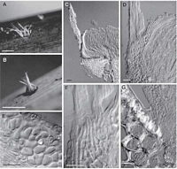
Caption: Fig. 5 Hispidula pounamu (A-C, PDD 70961; D-G, PDD 70962). A, B, macroscopic
appearance of apothecia; C, apothecium in vertical section (one side only); D, deta | 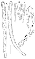
Caption: Fig. 6 Hispidula
pounamu (PDD 70961). A, asci; B, tips of paraphyses;
C, released ascospores; D, distribution
of collections examined. Scale bar: A-C = 20 µm. |
Article: Johnston, P.R. (2003). Hispidula gen. nov. (Helotiales, Hyaloscyphaceae) in Australia and New Zealand. New Zealand Journal of Botany 41(4): 685-697 (http://www.rsnz.org/publish/abstracts.php).
Description: DESCRIPTION: Apothecia developing on upper and lower surface of fallen leaves, typically
near base of leaf. Apothecia erumpent from beneath epidermis, arising from small patch of
compact, hyaline tissue comprising cylindric to short-cylindric, 2-5-µm diam.cells with walls
hyaline, thin, nongelatinous. Apothecia up to 1 mm diam., sessile, disc bright orange-yellow,
receptacle white to bright yellow, dark brown with age or drying, with a row of
massive, tapering, pale, whitish appendages up to 0.8 mm long. Ectal excipulum two-layered;
outer layer up to 50 µm thick, forming a broad, flange-like extension above the surrounding
host tissue, of textura angularis to textura prismatica with elements oriented at high angle to
receptacle surface, comprising brick-shaped, 8-12-µm-diam. cells with walls thick, hyaline,
gelatinous, outermost cells encrusted with yellow-brown material; inner layer 5-10 µm thick,
comprising narrow-cylindric to cylindric, 2-2.5-µm-diam. cells with walls thick, hyaline,
gelatinous. Medullary excipulum comprising 50-60-µm-deep layer of cylindric 3-5-µm-diam.
cells partly interwoven and oriented more or less perpendicular to host surface, with walls
thick, hyaline, gelatinous. Excipular hairs arising from outer layer of ectal excipulum, up to
800 x 3 µm, cylindric with rounded apex, walls thick, smooth, dextrinoid in Melzer's reagent
(under interference contrast microscopy with amyloid appearance at some angles), numerous
hairs aggregated into tight groups, forming triangular, tapering appendages, 40-80 µm wide at
base. Subhymenium comprising 2-µm-diam. hyphae with walls hyaline, thin,
nongelatinous. Paraphyses 1.5 µm diam., apex slightly swollen to 2.5-3.5 µm diam.,
sometimes branched in upper 20-40 µm, extending 20-30 µm beyond asci, embedded in
common gelatinous matrix. Asci (95)110-135 x 7-9.5 µm, cylindric, tapering slightly
to subtruncate apex, wall thickened at apex, apical pore amyloid, 8-spored, spores overlapping
uniseriate, spores extending 80-110 µm from ascus apex. Ascospores 13.5-19(-22) x 4-5.5
µm, oblong elliptic,ends rounded, flattened one side, slightly curved in side view, 0-1-septate,
wall hyaline (pale brown in some spores after release).
Notes: ETYMOLOGY: The specific epithet refers to the Maori name for the South Island of New
Zealand, Te Wai Pounamu, reflecting the largely southern distribution of this species (epithet
in the form of a noun inaposition).
NOTES: H. pounamu is similar to H. tokerau; see notes under this second species. H.
pounamu is quite common in the northern half of the South Island, the only North Island
collection being from the summit of Mt Te Aroha.
|