|
 Corticium utriculicum Corticium utriculicum
BiostatusPresent in region - Indigenous
Images (click to enlarge)
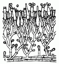
Caption: TEXT-FIG. 4. Corticium utriculicum. x 500. | 
Caption: 14. C. utriculicum, cystidia, stages in basidium development, spores. |
Article: Cunningham, G.H. (1954). Thelephoraceae of New Zealand. Part III: the genus Corticium. Transactions of the Royal Society of New Zealand 82(2): 271-327.
Description: Hymenophore annual, adnate, membranous, effused, forming irregular linear areas to 15 x 4
cm.; surface white, becoming cream, pruinose, creviced when old; margin thinning out,
arachnoid, white, adnate. Context white, 70-140 µ thick, composed of a narrow base of
parallel hyphae and an intermediate layer of scanty upright hyphae branched more freely
beneath the hymenium, with or without crystals; generative hyphae 3-4 µ diameter, wall 0.2 µ
thick, hyaline, branched, septate, with clamp comlections, sometimes crystal coated- near the
base. Hymenial layer to 50 µ deep, of basidia, paraphyses and gloeocystidia. Basidia
subclavate, subcylindrical, some constricted in the middle region, 16-24 x 4-5 µ, 4-spored,
projecting; sterigmata slender, to 6 µ long. Paraphyses subclavate or cylindrical, shorter and
narrower than the basidia. Gloeocystidia confined to the hymenial region, fusiform or
ventricose with acute apices, some capitate, 20-32 x 4-5 µ, projecting to 15 µ, or not, wall
0.25 µ thick. Vesicles arising from hyphae of the intermediate layer on short lateral
branchlets, pyriform or abruptly capitate, 4-5 µ diameter, smooth, staining deeply. Spores
oval, a few subglobose, 4-6 x 3-3.5 µ, wall smooth, hyaline, 0.2 µ thick.
Habitat: HABITAT. Effused on bark of decaying branches.
Distribution: DISTRIBUTION. New Zealand.
Notes: Specific characters are the fusiform or ventricose small gloeocystidia, small oval spores, and
lateral. pyriform vesicles. Gloeocystidia are confined to the hymenial region, apices being
usually long-acuminate and projecting slightly, though some are capitate. Vesicles arise from
lateral branchlets of the upright hyphae of the intermediate layer; they may be abundant, then
appearing in whorls on the hyphae, or scanty when but one or two may be seen near the base.
In old specimens tissues may collapse and become somewhat pseudoparenchymatous.
Crystals may be present or absent, consequently they have not been included in the drawing.
Article: Stalpers, J.A. (1985). Type studies of the species of Corticium described by G.H. Cunningham. New Zealand Journal of Botany 23(2): 301-310 (http://www.rsnz.org/publish/abstracts.php).
Description: Basidiome effused, membranaceous, slightly cracked. Hymenial surface even, cream-coloured; margin white, hypochnoid. Subicular hyphae hyaline, thin-walled, 2.5-4 µm wide,
sometimes slightly sinuous, with clamps. Cystidia hyaline, thin-walled, typically acuminate,
rarely capitate, basally inflated, sometimes with 1-3 constrictions (not apical), 1830 X 3.5-5
µm. Basidia suburniform, 18-25 X 4-5 µm. Spores hyaline, smooth, thin- to slightly thick-walled, broadly ellipsoid, 4.5-6 X 3.5-4.5 µm, not amyloid.
Notes: The species is close to Hyphoderma sambuci (Pers.) Rilich, a species which is intermediate
between Grandinia and Hyphoderma and therefore sometimes placed in Lyomyces P. Karst.;
the cultural characteristics of H. sambuci (Stalpers 1978) suggest Grandinia. Vesicles as
described by Cunningham were not observed. The study of additional material is necessary
before the status of the name can be established.
|