|
 Cordierites acanthophora Cordierites acanthophora
BiostatusPresent in region - Indigenous. Endemic
Images (click to enlarge)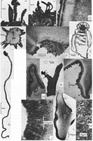
Caption: Fig. 1: A-C. Cordierites acanthophora: A. Apothecia as found in nature. - B. Apothecia on long stipe. -
C. Transverse section through stipe showing
peripherally situated pycnidia. (PDD 46956). -
D-G. Cryptohymenium pycnidiophor | 
Caption: Fig. 2: A-D. Cordierites acanthophora: A. Section through apothecium showing margin and ectal excipulum. - B. Asci and paraphyses. - C. Two ascospores. -
D. Phialides and conidia. (A: Holotype, B-D: PDD 46956).
E, F. Sclerocrana a |
Article: Samuels, G.J.; Kohn, L.M. (1987) [1986]. Ascomycetes of New Zealand. 7. Some bizarre, inoperculate discomycetes. Sydowia 39: 202-216.
Description: Apothecia 2-8 cm high, produced from decaying herbaceous debris, scattered, consisting of a
cylindrical stalk bearing thorn-like pycnidial projections and 2-3
apical, and 1-2 opposed, intercalary 3-4 mm diam. discs. Hymenium dark brown, becoming nearly
black when dry; fringed with regularly-spaced, flat, triangular,
dark brown teeth; when fresh flat; receptacle dark brown, surface rugose. Stipe cylindrical,
in fresh condition yellow brown, becoming dark brown with age and
when dry; surface with coarse warts and scattered pycnidia that appear as acute, dark brown to
black thorns ca. 1.5 mm high; terminating in many coarse,
unbranched, dark brown to black hyphae.
Asci 8-spored, (175-)190-223(-239) x (7-)9-10 µm, cylindrical, base forming a
slight foot, wall ca. 1 µm thick; pore J + Melzer's, cylindrical, ca. 3 µm long x 3
µm wide; ascospores forming in the apical ca. 120 µm of the ascus. - Ascospores (16-)22-28.8(-38) x
(7-)7.6-9(-10) µm, obliquely uniseriate with overlapping
ends; at first hyaline, becoming light brown within the ascus, fusiform with subacute to acute ends,
with scattered, 1-1.5 µm diam. warts. Paraphyses 10-20 µm
longer than asci, consisting of a ca. 3 µm wide stalk and a clavate to subglobose, 7-10 µm wide
tip; hyaline, unbranched or infrequently branched near the apex,
septate, cells 20-40 µm long; tips of paraphyses encrusted in amorphous brown material, forming a
pseudoepithecium. - Subhymenium 50-80 µm wide, not
staining in Melzer's reagent, consisting of compacted, intertwined, 3-8 µm wide, smooth, light brown
hyphae; depositions of brown pigment scattered in the
subhymenium. Medullary excipulum well developed, not delimited from the ectal excipulum, not staining
in Melzer's reagent, consisting of tightly compacted,
nearly circular, 10-15 µm diam. cells with walls ca. 1.5 µm thick, immediately below the subhymenium;
cells of medullary excipulum gradually becoming
vertically elongated, 20-40 x 10-15 µm with walls ca. 1.5 µm thick and merging with the cells of the
stipe. Ectal excipulum consisting of a single region of cells
125-200 µm wide along flanks, narrowing to ca. 60 µm along the stipe, consisting of textura angularis,
cells 25-50 µm across, walls ca. 1.5 µm thick, light
brown; continuing outward ± perpendicular to surface of receptacle to form triangular teeth consisting
of cingular cells with long axis ± perpendicular to surface
of receptacle cells 15-30 x 8-20 µm, walls ca. 1.5 µm thick, brown. - Stipe with sharply delimited,
ca. 125 µm wide cortex consisting of vertically oriented
hyphal cells 16-25 x ca. 15 µm, walls 1.5-3.5 µm wide brown; cells at surface of stipe joined into
scattered warts; medulla of stipe consisting of vertically
oriented, hyphal cell, 30-40 µm x 5-10 µm wide with walls 1 µm thick, non-pigmented; stipe circular
in cross section. Pycnidia arising along length of stipe, cells
at surface of stipe continuous with cells at surface of pycnidium; cells of medulla of stipe
continuous with internal tissue of pycnidium; wall of pycnidium ca. 80
µm wide; conidiogenous cells arising directly from cells of the inside of the pycnidial wall,
11-15 x 3-4 µm at base tapering ± uniformly to 1-2 µm wide at tip,
sometimes widest in the middle, phialidic, monoblastic, pale brown. Conidia 3.5-5.0 x 2-3 µm,
oblong, lacking a basal abscission scar, unicellular, hyaline.
Habitat: Growing in soil at base of Leptospermum sp. and in decaying wood (? Leptospermum).
Notes: On the basis of gross morphology Cordierites acanthophora can easily be
accommodated in the genus Cordierites. Whether the species is actually
congeneric with other species of the genus is, however, questionable. Apothecial tissues of
species of Cordierites (sensu KORF 1973) are ionomidotic, but
apothecial tissues of C. acanthophora are not ionomidotic. The large brown, warted
ascospores are unprecedented in Cordierites but they are also unusual
among the inoperculate discomycetes and give no clues as to the affinities of the species.
The ectal excipulum comprising dark angular cells and the pycnidial
anamorph argue for the inclcusion of the species in the Dermateaceae rather than the
Leotiaceae, where KORF (1973) placed Cordierites. Cordierites, however,
is a poorly known genus and in the absence of a critical study of its species, we prefer
to refer C. acanthophora to it.
|