|
 Coccomyces longwoodicus Coccomyces longwoodicus
BiostatusPresent in region - Indigenous. Endemic
Images (click to enlarge)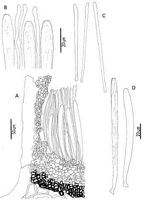
Caption: Fig. 13 Coccomyces longwoodicus: A, ascocarp margin in cross-section. B, apices of asci and
paraphyses. C, released ascospores. D, asci. | 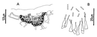
Caption: Fig. 14 Anamorph of Coccomyces longwoodicus: A, pycnidium in cross-section. B,
conidiogenous cells and conidia. | 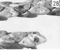
Caption: Fig. 28 Macroscopic appearance of ascocarp (x 12).
C. longwoodicus |
Article: Johnston, P.R. (1986). Rhytismataceae in New Zealand. 1. Some foliicolous species of Coccomyces de Notaris and Propolis (Fries) Corda. New Zealand Journal of Botany 24(1): 89-124 (http://www.rsnz.org/publish/abstracts.php).
Description: Ascocarps forming within pale, brownish lesions on fallen leaves. Lesions are not associated
with zone lines. Ascocarps irregularly oblong to ovate in outline, 0.7-1 mm diam. When
immature walls pale grey to. concolorous, darker around the outside edge, with no obvious
paler zones along future lines of opening. Mature ascocarps opening by irregular, radiate
splits, with the covering tissue sometimes eroding to widely expose the yellow hymenium.
Pycnidia present, flat, lenticular, pale brown, darker around the edge, containing conidia only
when associated with immature ascocarps.
Ascocarps initially subcuticular, with some epidermal cells becoming filled with fungal tissue
as the ascocarp ages. In vertical section upper stromatal layer 15-20 µm wide, comprising
brown to pale brown, thin walled, rounded cells, 4-6 µm diam. This layer does not extend as
far as the outer edge of the ascocarp. The area between the upper and lower stromatal layers is
filled with hyaline, gelatinous tissue. Cylindric, vertically orientated, hyaline, gelatinous-walled cells develop between the upper stromatal layer and the covering host tissue. These
cells form a well-developed layer in immature ascocarps, but are often difficult to distinguish
after the ascocarp has opened. A few periphyses, 12-17 x 2-3 µm, line the inside of the
upper wall. The lower stromatal layer is 60-70 µm wide; the outer 30-40 µm is composed
of dark brown, thick walled, globose cells, 5-8 µm diam., the inner part composed of
hyaline, globose cells, 6-10 µm diam. Subhymenium 10- 15 µm wide, of hyaline,
gelatinous-walled cells.
Paraphyses 2-2.5 µm diam., gradually increasing in width to 3-4 µm diam. at the apex, not
branching, not gelled together, extending 20-30 µm beyond the asci. Asci 137-155 x 8.5-10
µm, subclavate, tapering in slightly to the broadly truncate to slightly rounded apex, the wall
sometimes with a short, broad apical pore, non-amyloid, 8-spored. Ascospores 58-72 x 2.5-3
µm, tapering slightly to basal end with a small swelling at the very base, 0-septate, straight
when released, gelatinous cap at both ends.
Pycnidia intraepidermal, flat, lenticular in shape, lower wall of brown, angular cells, upper
wall absent. Conidiogenous layer lining the lower wall, comprising a palisade of solitary,
cylindric, hyaline, sympodial conidiogenous cells, 8-16 x 2-3 µm. Intermixed amongst the
conidiogenous cells, especially near the centre of the pycnidia are filiform, hyaline, sterile
elements, 45-60 x 1-2 µm. Conidia, 5-6 x 1-2 µm, short-cylindric, rounded at both ends,
hyaline, 0-septate.
Habitat: Has been found only on Halocarpus biformis.
Notes: ETYMOLOGY: longwoodicus; refers to type locality.
NOTES: The ascocarps of C. longwoodicus are similar in macroscopic appearance to two
other species found on southern conifers, C. cupressinum and C. libocedri, with all having a
pale, poorly developed upper ascocarp wall. The three species can be distinguished using
ascus and ascospore size.
|