|
 Coccomyces globosus Coccomyces globosus
BiostatusPresent in region - Indigenous. Non endemic
Images (click to enlarge)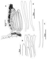
Caption: Fig. 6 Coccomyces globosus: A, ascocarp margin in cross-section. B, apices of asci and
paraphyses. C, released ascospores. D, asci. | 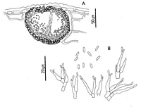
Caption: Fig. 7. Anamorph of Coccomyces globosus: A, pycnidium in cross-section. B, conidiogenous
cells and conidia. | 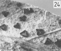
Caption: Fig. 24 Macroscopic appearance of ascocarp (x 12).
C. globosus; | 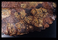
Caption: Kaimanama, Clements Mill Rd. 4/2/89
Owner: Herb. PDD | |
Article: Johnston, P.R. (1986). Rhytismataceae in New Zealand. 1. Some foliicolous species of Coccomyces de Notaris and Propolis (Fries) Corda. New Zealand Journal of Botany 24(1): 89-124 (http://www.rsnz.org/publish/abstracts.php).
Description: Ascocarps usually developing on the lower leaf surface, within pale, yellowish or brownish
lesions, not associated with narrow, black zone lines but dark brown, broad, diffuse-edged
zone lines present around lesions on Weinmannia racemosa. Ascocarps angular in outline, 3-4(-5) sided, 0.5-0.8 mm diam. Immature ascocarps with walls pale grey to black with faint to
well developed paler zones along future lines of opening, and often with a paler zone just
inside edges of ascocarp. Pale grey ascocarps delimited along outside edge by narrow, black
line. Mature ascocarps open by 3-5 radiate slits, walls pale brown to black, sometimes with a
paler zone adjacent to outer edge. No lip cells. Pycnidia always present, globose to depressed-globose, immersed, black walled, paler near centre.
Ascocarps intraepidermal. In vertical section upper stromatal layer 30-50 µm wide,
comprising pale brown to brown, thin to thick walled, more or less globose cells, 4-8 µm
diam. Cells becoming paler and thinner walled near outer edge of ascocarp. Periphyses
absent. Lower stromatal layer joining the upper, 15-25 µm wide, of 4-5 layers of dark
brown, thick walled, more or less globose cells, 5-9 µm diam., the outer layer of cells with
darker and thicker walls. Subhymenium 10-20 µm wide, of hyaline, thin walled, rounded
cells, 3-8 µm diam. Layer of disorganised hyphal tissue, 20-50 µm wide, between
subhymenium and lower wall. Excipulum poorly developed, comprising a few marginal
paraphyses becoming closely septate and sometimes capped by dark brown material.
Paraphyses 2-2.2 µm diam., swollen to 4-7 µm diam. at the clavate or fusoid apices,
extending 20-30 µm beyond asci. Asci 104-140 x (5-)5.5-7(7.5) µm, cylindric to
subclavate, tapering slightly to rounded apex, non-amyloid, 8-spored. Ascus apical thickening
and broad pore sometimes seen when immature. Ascospores filiform, 55-78 x 1.2-1.8 µm
tapering slightly to basal end, more or less straight when released, 0-1 septate, gelatinous
sheath present (see notes below).
Pycnidia depressed-globose, intraepidermal to subepidermal, ostiole not well-defined, often
opening through a stomatal cell. Pycnidial wall continuous, not divided into upper and lower
parts, 5-12 µm wide, of 1-3 layers of brown, somewhat flattened, pseudoparenchymatous
cells, 2-3.5 µm diam. Conidiogenous layer lining whole pycnidium, comprising hyaline,
flask-shaped, sympodial conidiogenous cells, 9-18 x 1.5-3.8 µm, with a long, flexuous neck,
often with two conidia held at the ends. Conidiogenous cells solitary or borne on poorly
developed, irregularly branching conidiophores, intermixed with filiform, sterile elements,
especially in area beneath ostiole. The sterile elements may form a column of tissue, almost
reaching to the top of the pycnidium. Conidia short-cylindric, rounded ends, 0-septate,
hyaline, (3.8-)4.5-6 x 0.8-1(-1.2) µm.
CHARACTERISTICS IN CULTURE: Ascospores of PDD 43034 and PDD 43964
germinated within 5-6 days. Cultures on oatmeal agar 3.5-4 cm diam. after 4 weeks; aerial
mycelium sparse, felted, white; agar surface grey toward centre of colony, yellowish toward
edge; reverse, yellow. Pycnidia forming near centre of colony, globose, black walled; conidial
ooze yellow. Conidiogenous cells and conidia of same appearance and size as those found in
nature.
Habitat: Found on fallen leaves of Nestegis lanceolata, Weinmannia racemosa, and
Metrosideros fulgens.
Notes: ETYMOLOGY: globosus = globose, spherical; refers to shape of anamorph pycnidium.
NOTES: Some of the collections on Weinmannia racemosa (PDD 44649, 44650, 44653,
44928, 44962) can be separated from typical C. globosus in having asci 7-9 µm wide, and
broadly truncate at the apices, and ascospores (71-)80-98 µm long. In all other respects,
including the anamorph, they are identical to C. globosus. They have been included here in C.
globosus.
The anamorph pycnidium of C. globosus is unusual when compared to those of most other
Rhytismataceae, in not being flattened, and in having the conidiogenous layer lining the
whole pycnidial cavity. It is similar to that described for Lophodermium camelliicola by
Minter & Sharma (1982).
C. globosus is similar in hymenial dimensions to C. phyllocladi. C. phyllocladi differs in that
it lacks pycnidia, and has an excipulum which forms from a layer of gelatinised tissue
between the hymenium and the outer stromatal layers. Cultural characteristics also differ.
C globosus is also similar to C. antillarum Sherwood and C. spegazzinii Saccardo. From
Sherwood's 1980 descriptions, C. antillarum and C. spegazzinii are more or less the same in
ascus and ascospore size and shape, and ascocarp margin structure. They appear to differ only
in C antillarum lacking zone lines around the lesions, having an excipulum which remains
hyaline, and having periphyses lining the upper stromatal wall. The specimens of C
antillarum studied (R. Thaxter, 1912-13, on unidentified leaves, Grand Etang, Grenada -
holotype FH; Pfister 1006, on unidentified leaves, Grand Etang, Pfister, Carpenter, Sherwood
6.1.1974 - FH) agreed well with the description of Sherwood (1980), but the only specimens
of C. spegazzinii examined (Brasil, Apiahy, Puiggari, 1889 - LPS 34990; Venezuela, Edo.
Sucre, Dumont et al., 15.VII.1972, NY-VE5083) were small and in poor condition, and did
not match the description of the hymenium of C. spegazzinii given by Sherwood. Compared
with C. globosus these species have the same macroscopic appearance and hymenial element
dimensions, and the excipulum and the way it develops are also similar. C globosus can be
separated from C. antillarum in having an anamorph, and in lacking periphyses. C spegazzinii
is apparently associated with zone lines, and lacks an anamorph. However, considering the
small number of collections of C. antillarum and C. spegazzinii in herbaria and the relatively
minor differences between all three taxa, they may in fact be found to be conspecific when
more South American collections are available for comparison.
Article: Gadgil, P.D. (in association with Dick, M.A.; Hood, I.A.; Pennycook, S.R.) (2005). Fungi on trees and shrubs in New Zealand. Fungi of New Zealand. Ngā Harore o Aotearoa 4: xi + 437 p. Hong Kong: Fungal Diversity Press.
Description: Type: Foliicolous Fungi; Description: Ascomata apothecial, scattered, intraepidermal, angularly 3- to 5-sided, up to 1 mm in diameter, black, opening by several radial splits; on discrete, creamy brown to yellow brown areas up to 10 mm across, on both sides of necrotic or fallen leaves. Asci cylindric to subclavate, 95–120 × 5–7 μm. Ascospores filiform, 0-septate, 50–75 × 1.5–2 μm, hyaline. Conidiomata scattered among the ascomata, pycnidial, submerged, depressed globose, black, 0.1–0.13 mm in diameter, with an ostiole located beneath a stoma. Conidia short cylindrical, 0-septate, 4–6 × 1 μm, ends obtuse.
Distribution: Distribution: Auckland, Coromandel, Taranaki, Taupo, Gisborne, Nelson, Buller, Westland.; 1st Record: Johnston (1986).
|