|
 Coccomyces crystalligerus Coccomyces crystalligerus
BiostatusPresent in region - Indigenous. Non endemic
Images (click to enlarge)
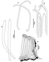
Caption: Fig. 2. Coccomyces crystalligerus: A, ascocarp margin in cross-section. B, apices of asci and
paraphyses. C, released ascospores. D, asci. | 
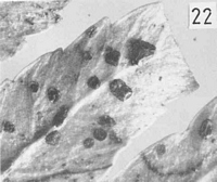
Caption: Fig. 22 Macroscopic appearance of ascocarp (x 12).
C. crystalligerus; | 
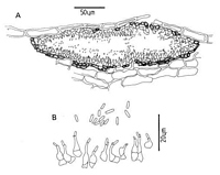
Caption: Fig. 3 Anamorph of Coccomyces crystalligerus: A, pycnidium in cross-section. B,
conidiogenous cells and conidia. | 
Owner: Herb. PDD | |
Article: Johnston, P.R. (1986). Rhytismataceae in New Zealand. 1. Some foliicolous species of Coccomyces de Notaris and Propolis (Fries) Corda. New Zealand Journal of Botany 24(1): 89-124 (http://www.rsnz.org/publish/abstracts.php).
Description: Ascocarps developing on lower frond surface, in pale, brownish lesions, not associated with
zone lines. Ascocarps more or less round in outline, 0.3-0.6 mm diam. Immature with walls
pale to dark grey with a well-developed, longitudinal or branching, broad, pale zone along
future line of opening. Edge of ascocarp marked by a narrow, black line. Mature ascocarp
steep-sided, walls pale grey, black along outer edge of ascocarp and along edges of opening.
Shape of opening irregular, scalloped. Pycnidia present on most collections, round to oblong
in outline, variable in size up to 0.3 x 0.5 mm, pale brown, darker at the edge and around the
one to several, round ostiolata.
Ascocarps subepidermal. In vertical section upper stromatal layer poorly developed, 20-25
µm wide, not becoming wider around opening, composed of brown to very pale brown, thick
walled, angular to globose cells, 3-6 µm diam. Inside of stromatal layer lined with periphyses
when immature. Excipulum becoming well-developed as ascocarps mature, arising from inner
layers of upper stroma, comprising hyaline, closely septate hyphae, which become swollen,
and embedded in dark brown material at apices. Lower stromatal layer 5-15 µm wide, the
outer 1-2 layers of cells dark brown and thick walled, with hyaline, globose cells to the inside.
Paraphyses 1-1.5 µm diam., branching several times near apices not or only slightly swollen.
intermixed with crystals, extending 15-20 µm beyond asci. Asci cylindric to subclavate, (132-)150-195(-210) x 7.5-11 µm, tapering to an acute apex, wall not thickened at apex, non-amyloid, 8-spored. Ascospores filiform, (95-)110-125 x 1.8-2.5 µm, tapering slightly to both
ends, 0-1 septate, often coiling when released, surrounded by poorly developed gelatinous
sheath.
Pycnidia subepidermal. In vertical section lenticular in shape, wall 5-10 µm wide, of brown,
angular cells, lined with hyaline, thin walled cells on which the conidiogenous layer is held.
The lower wall is wider and darker than the upper. Conidiogenous cells lining both upper and
lower walls, comprising flask-shaped, sympodial, solitary conidiogenous cells, often with two
conidia held at the ends. Conidia short-cylindric, 3.5-5.5 x 1 µm, hyaline, non-septate.
Habitat: Found on fronds of ferns, including Dicksonia, Blechnum, and Asplenium species.
Although the ascocarps are always found on dead tissue, they are sometimes in a small lesion
on an otherwise apparently healthy leaf
Notes: The ascocarps of the type specimen of C. crystalligerus are larger, have a rounder,
more regular-shaped opening, and better developed layer of crystals on the hymenium,
compared with New Zealand collections. Pycnidia are not present on the type specimen, but
were found on some, but not all, of the New Zealand collections. Otherwise the New Zealand
collections of this fungus fit the description of the type specimen well, especially with regard
to the host, the appearance and size of the hymenial elements, and the structure of the
ascocarp margin.
The only other rhytismataceous taxon so far consistently found on ferns in New
Zealand is a Hypoderma-like species on Gleichenia cunninghamii Heward ex Hook.
C. radiatus was found once on Dicksonia squarrosa
|