|
 Simocybe phlebophora Simocybe phlebophora
BiostatusPresent in region - Indigenous. Endemic
Images (click to enlarge)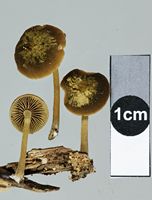
Owner: J.A. Cooper | 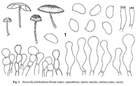 | 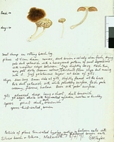
Caption: Watercolour
Owner: G.M. Taylor | 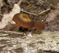
Owner: J.A. Cooper | 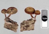
Owner: J.A. Cooper | 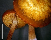
Caption: scale=5mm
Owner: J.A. Cooper | 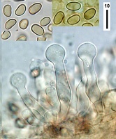
Caption: spores and cheilocystidia
Owner: J.A. Cooper | 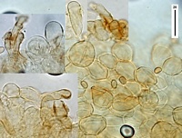
Owner: J.A. Cooper | 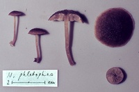
Caption: ZT67-174 , Holotype
Owner: E. Horak: © Creative Commons Attribution-Noncommercial 3.0 New Zealand | 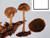
Owner: J.A. Cooper | 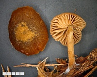
Owner: J.A. Cooper | 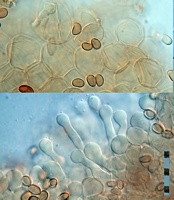
Caption: top: cap cells. bottom: cheilocystidia.
Owner: J.A. Cooper | 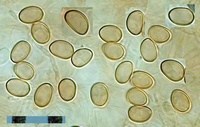
Caption: spores
Owner: J.A. Cooper | 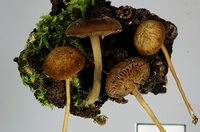
Owner: J.A. Cooper | 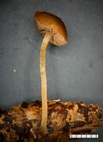
Owner: J.A. Cooper | 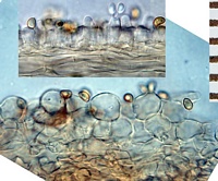
Caption: top: gill section with basidia. Bottom: section through cap surface.
Owner: J.A. Cooper | 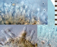
Caption: cheilocystidia
Owner: J.A. Cooper | 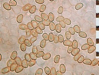
Caption: spores
Owner: J.A. Cooper | 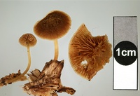
Owner: J.A. Cooper | 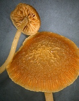
Owner: J.A. Cooper | 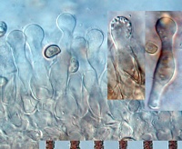
Caption: cheilocystidia (KOH)
Owner: J.A. Cooper | 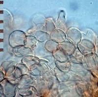
Caption: section through cap surface (KOH)
Owner: J.A. Cooper | 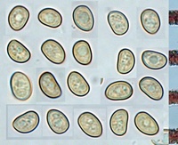
Caption: spores (KOH)
Owner: J.A. Cooper | 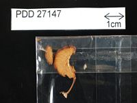
Caption: Dried type specimen
Owner: Herb PDD | |
Article: Horak, E. (1980). Fungi Agaricini Novazelandiae. X. Simocybe Karsten. New Zealand Journal of Botany 18(2): 189–196 (http://www.rsnz.org/publish/abstracts.php).
Description: Pileus -30 mm, hemispheric to convex, becoming plane; grey-brown to dark brown with distinct olive tint; dry, coarsely wrinkled to venose (at least at disc), net-like structure usually very conspicuous, striate towards margin, strongly hygrophanous, veil remnants none. Lamellae (L 10-14, -3) crowded, adnexed or adnate becoming emarginate with age, ventricose; pale argillaceous turning brown, edge albofimbriate. Stipe -30 x -2.5 mm, cylindric, equal, central; pale brown, paler at apex; dry, pruinose above, longitudinally fibrillose towards white. villous base, solid, veil remnants lacking, single (and cespitose) in groups. Context brown with olive tint. Odour and taste acidulous. Spore print brown. Spores 6-7.5 x 4-5 µm, broadly phaseoliform, membrane thin-walled, brown, smooth, germ pore none. Basidia 15-25 x 5-6 µm, 4-spored. Cheilocystidia 25-40 x 8-13 µm, fusoid with broad, capitate apex, membrane thin-walled, hyaline, pigment absent. Pleurocystidia absent. Caulocystidia like cheilocystidia but larger. Cuticle a celluloderm of erect chains of ovoid to globose cells (10-30 x 12-20 µm), membrane not gelatinised, encrusted with brownish (KOH) pigment. Clamp connections present.
Habitat: o n rotten wood of Nothofagus (N. menziesii (Hook.f.) Oerst., N. solandri (Hook.f.) Oerst. var. cliffortioides (Hook.f.) Poole). New Zealand.
Notes: The conspicuous vein-like net on the brown-olive pileus is a diagnostic feature not found among the other known species of New Zealand Simocybe. S. phlebophora is further characterised by the cellular structure of the pileocuticle and the fusoid-capitate cheilo- and caulocystidia.
|