|
 Coccomyces clavatus Coccomyces clavatus
BiostatusPresent in region - Indigenous. Non endemic
Images (click to enlarge)
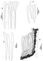
Caption: Fig. 1. Coccomyces clavatus: A, ascocarp margin in cross-section, B, apices of asci and
paraphyses. C, released ascospores. D, asci. | 
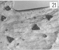
Caption: Fig. 21 Coccomyces clavatus; | 
Caption: Type
Owner: Herb. PDD |
Article: Johnston, P.R. (1986). Rhytismataceae in New Zealand. 1. Some foliicolous species of Coccomyces de Notaris and Propolis (Fries) Corda. New Zealand Journal of Botany 24(1): 89-124 (http://www.rsnz.org/publish/abstracts.php).
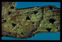
Description: Ascocarps forming within pale, yellowish lesions on fallen leaves. Lesions not associated with
zone lines. Ascocarps angular, 3-4 sided, 0.6-0.8 mm diam. Immature with walls pale grey to
black, with paler zones along future lines of opening. Mature with walls pale grey to black,
opening widely to expose the yellow hymenium. Lip cells absent. Pycnidia absent.
Ascocarps intraepidermal. In vertical section upper stromatal layer 15-30 µm wide,
comprising brown to dark brown, angular cells, 5-8 .µm diam. A few cylindric, hyaline,
periphysis-like cells, 10-15 x 2-3 µm, near opening. Lower stromatal layer 20-30 µm wide, of
3-5 layers of dark brown, thick walled cells 5-8 µm diam. A 15-20 µm wide layer of
gelatinised tissue present between the lower stromatal wall and the sub-hymenium, extending a
short distance up the sides of the hymenium, giving rise to few, branching, excipular-like
elements.
Paraphyses 1.5-2 µm diam., swollen to 4-6 µm at the clavate apices, extending 10- 15 µm
beyond asci. Asci 101-118 x 7.5-9 µm, subclavate, tapering in to a broadly truncate apex,
wall uniform in thickness, non-amyloid, 8-spored. Ascospores 35-43.5 x 2-3 µm, clavate,
tapering to the base, 0(-1) septate, hyaline, with a gelatinous cap.
Habitat: Fallen phylloclades of Phyllocladus aspleniifolius var. alpinus.
Notes: ETYMOLOGY: clavatus = clavate; refers to ascospore shape.
C. clavatus is similar to C. phyllocladi, also found on Phyllocladus. The two species
are indistinguishable macroscopically, and are similar in appearance in cross-section, both
having a characteristic, gelatinous layer between the lower stromatal wall and the sub-hymenium. They differ in the size and shape of the asci and ascospores.
No rhytismataceous species have previously been reported on Phyllocladaceae. Two
Hypoderma-like species have also been found on this host in New Zealand.
|