|
 Rectipilus fasciculatus Rectipilus fasciculatus
SynonymsLachnella fasciculata
Solenia candida
Solenia fasciculata
BiostatusPresent in region - Indigenous. Non endemic
Images (click to enlarge)
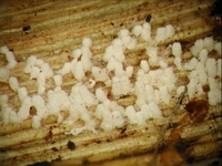
Caption: fruitbodies
Owner: J.A. Cooper |
Article: Cunningham, G.H. (1963). The Thelephoraceae of Australia and New Zealand. New Zealand Department of Scientific and Industrial Research, Bulletin 145: 359 p. Wellington:.
Description: Subiculum annual, arachnoid,
white, effused forming areas to 10 cm across. Pilei densely aggregated,
sometimes confluent, seldom scattered, cylindrical or subclavate, 0.5-3 mm tall,
0.1 -0.4 mm diameter, attached by narrow bases, ceraceous, brittle, white drying
honey yellow; pileus surface covered with tortuous encrusted (in part)
abhymenial hairs; margin even, inrolled and slightly thickened. Context white,
to 35 µm thick, of densely compacted parallel hyphae; generative hyphae 2.5-3 µm
diameter, walls 0.2 µm thick, hyaline. Hymenial layer to 30 µm deep, a close
palisade of basidia and paraphyses. Basidia clavate, 12-16 x 5-6 µm, bearing 4
spores; sterigmata erect, slender, to 4 µm long. Paraphyses subclavate, 10-14 x
4-5 µm. Spores subglobose, a few globose or oval, 4.5-5.5 µm diameter, walls
smooth, hyaline, 0.2 µm thick.
Habitat: HABITAT: Crowded
often in fascicles on bark or decorticated dead branches or dead stipes of tree
ferns.
Distribution: DISTRIBUTION: Europe, Great Britain, North and South
America, Australia, New Zealand.
Notes: Pilei
grow in dense clusters, rarely solitary. At first globose, they soon become
cylindrical. The exterior is covered with tortuous, aseptate, simple abhymenial
hairs which later may disappear though some may remain near apices. The
subiculum is arachnoid, delicate and often difficult to detect unless specimens
are examined under a dissecting microscope. Spores vary in size and shape; when
first formed, walls are relatively thick, but as spores mature walls become
thinner. In a previous paper (1953b, p. 176) I listed "Solenia" fasciculata
as a synonym of S. candida . W. B. Cooke has advised that the two
may be separated by the abhymenial hairs, those of the former being filiform and
encrusted, of S. candida delicate, naked, and branched near apices. In
other particulars they appear to be similar, save that pilei of L. candida
are seldom crowded into fascicles.
|