|
 Physalacria pseudotropica Physalacria pseudotropica
BiostatusPresent in region - Indigenous. Endemic
Images (click to enlarge)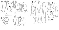
Caption: Physalacria pseudotropica (TNS-F-171685). A. Basidia and basidioles in hymenium. B. Basidiospores. C. Cells from sterile capitulum base. D. Hymenial gloeocystidia. E. Caulocystidia. Bars: A-E 10 µm. |
Article: Tanaka, I.; Doi, Y.; Hongo, T. (2004). Two unusual species of Physalacria (Basidiomycetes, Agaricales) collected in New Zealand and Papua New Guinea during mycological expeditions by the National Science Museum, Tokyo. Mycoscience 45(2): 143-146 Mycological Society of Japan.
Description: Basidiomata 0.7-3.0mm tall, stipitate-capitate. Capitulum 0.3-1.5mm diameter, hollow, globose to subglobose, pruinose, grayish-orange after drying. Resinous exudates on apex of hymenial cystidia were observed under the stereoscope. Stipe 0.4-2.0 X 0.06-0.08 mm, central, cylindrical, with pruinose cushion at the base. Basidiospores 3.8-5.9 X 2.3-3.4 um {x = 4.9 ± 0.5 X 2.9 ± 0.3 um; Q = 1.3-2.1; Q = 1.7 ± 0.2; n = 25), oblong to lacrymoid, smooth, hyaline, thin walled, inamyloid. Basidia 15-25 X 3.5-5.0 µm, narrowly clavate, 4-spored or rarely 2-spored; sterigmata 2.5-3.5 um long. Basidioles cylindrical to fusiform. Hymenial gloeocystidia 35-95 X 10-19 um, cylindrical to fusiform, obtuse or subcapitate, with resinous exudates adherent to the apex, projecting up to 35 um beyond hymenial cells; resinous exudates persist after drying, insoluble in Melzer's reagent, and gradually soluble in 3% KOH. Subhymenial hyphae 2-4 urn diameter, subagglutinate - to interwoven sparsely, cylindrical, hyaline, thin walled, inamyloid. Lower portion of capitulum composed of a hymeniform layer of fusiform to narrowly lageniform, sometimes rostrate cells; the cells hyaline, thin walled, inamyloid. Stipe tissue monomitic; cortical hyphae 2-4 µm diameter, parallel, cylindrical, smooth, hyaline, thin walled, inamyloid; medullary hyphae 2-6 um diameter, otherwise similar to cortical hyphae. Stipe base with scattered, curled, and thin to slightly thick walled (up to 0.8 um diameter) hyphal outgrowths up to 40 µm long and 1.5-3.0µm diameter. Caulocystidia abundant, conical to fusiform, subcapitate at the apex, with resinous exudates adherent to the apex; conical caulocystidia 17-37 X 12-23 um; fusiform caulocystidia 30-65 X 9-16 um. Clamp connections present on the hyphae in all tissues except some stipe tissues.
Habitat: Caespitose to solitary on decaying wood.
Distribution: New Zealand.
Notes: Specimens TNS-F-171685, -101517 accord well with P. pseudotropica in having a combination of small baisiospores (3.8-5.9 X 2.3-3.4nm), subcapitate hymenial gloeocystidia, and caulocystidia.
Physalacria pseudotropica was only reported from Gisborne (North Island) in New Zealand (Berthier 1985). In this report this fungus is recorded from South Island in New Zealand for the first time.
Resinous exudates of TNS-F-171685, -101517 were observed not only in water but also in Melzer's reagent and 3% KOH. Moreover, these were not soluble in water or Melzer's reagent. Persistence of resinous exudates in P. pseudotropica was not mentioned in the original description.
|