|
 Phlebia leptospermi Phlebia leptospermi
SynonymsCorticium leptospermi
BiostatusPresent in region - Indigenous. Endemic
Images (click to enlarge)
Caption: TEXT-FIG. 28. Corticium leptospermi. x 500. | 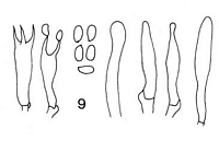
Caption: 9.C. leptospermi, basidia, spores and cystidia. |
Article: Cunningham, G.H. (1954). Thelephoraceae of New Zealand. Part III: the genus Corticium. Transactions of the Royal Society of New Zealand 82(2): 271-327.
Description: Hymenophore annual, adnate, ceraceous, effused, forming irregular areas to 10 x 5 cm.;
surface when fresh saffron- or chrome-yellow, drying buff or fawn, even, becoming deeply
areolately creviced, and tending to peel away in flakes; margin thinning out, adnate,
concolorous, vernicose when fresh. Context yellow, 100-200 µ thick, composed of a narrow
basal layer of parallel hyphae and an intermediate layer of compacted vertical hyphae coated
with yellow mucilage granules, masses of crystals embedded in lower portions; generative
hyphae 2.5-3 µ diameter, wall 0.2 µ thick, naked, branched, hyaline, septate, with abundant
clamp connections. Hymenial layer to 35 µ deep, of basidia, paraphyses and paraphysate
hyphae closely compacted. Basidia subclavate, 14-25 x 3-5 µ, not projecting, 2-4-spored;
sterigmata slender, to 6 µ long. Paraphyses cylindrical or subelavate, narrower than the
basidia. Paraphysate hyphae cylindrical or aculeate, abundant, projecting to 10 µ. Spores
broadly elliptical, 4-5.5 x 2.5-3 µ, abundant, wall smooth, hyaline, 0.2 µ thick.
Habitat: HABITAT. Effused on dead standing decorticated trunks.
Distribution: DISTRIBUTION. New Zealand.
Notes: Separated from other ceraceous species bearing mucilage by the chrome-yellow colour (when
fresh) of the deeply areolated surface, small basidia and presence of abundant paraphysate
hyphae projecting to 10 µ. Both context hyphae and tissues of the hymenium are coated with
fine granules of yellow mucilage, which gives colour to the plant; and the lower part of the
context contains additionally islands of crystals which may be scattered or arranged in the
form of lenses. Type specimens were taken from bases of fire-killed decorticated trunks on
which the fungus formed encircling sheets at or below the level of surrounding mosses.
In several features the species resembles the description of Peniophora sacchari Burt which
was recorded from Porto Rico on sugarcane trash; but is not so referred since type material
has not been examined.
Article: Stalpers, J.A. (1985). Type studies of the species of Corticium described by G.H. Cunningham. New Zealand Journal of Botany 23(2): 301-310 (http://www.rsnz.org/publish/abstracts.php).
Description: Hymenial surface even, chrome-yellow at the margin and more orange to reddish brown
towards the centre, turning red in KOH. Cystidia hyaline, thin-walled, clavate to capitate,
smooth or sometimes (in older parts) covered with resinous material, projecting up to 12 µm.
Spores hyaline, thin-walled, smooth, ellipsoid, 4.2-5 X 2-2.3 x 2.2-2.5 µm, not amyloid.
Notes: The species, which causes a weak white rot, belongs to Phlebia and is close to Ph.
femsjoeensis (Litsch. & Lund.) J. Erikss. & Hjortst., which has a distinct warted to phlebioid
hymenial surface and longer cystidia. It also resembles Ph. lilascens (Bourd.) J. Erikss. &
Hjortst., which has no cystidia and often a pinkish to violaceous colour.
|