|
 Clavaria megaspinosa Clavaria megaspinosa
BiostatusPresent in region - Indigenous. Endemic
Images (click to enlarge)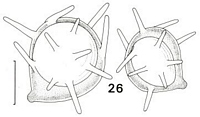
Caption: Fig 26. Clavaria megaspinosa. TENN no. 42418. Scale bar = 5 µm. | 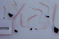
Caption: Microfiche 1-19. Clavaria megaspinosa. TENN no. 42418. |
Article: Petersen, R.H. (1988). The clavarioid fungi of New Zealand. New Zealand Department of Scientific and Industrial Research, Bulletin 236: 170 pp. Wellington:.
Description: Fruit bodies up to 50 x 2 mm, simple clubs, single or gregarious, slender, gracile, arising
from a small, white mycelial patch. Club equal, delicate coral-pink ("orange-pink", "pale
flesh color", "shrimp-pink", "grenadine-pink"), opaque to subtranslucent, appearing waxy;
flesh concolourous; apex narrowly rounded. Stipe up to 1 mm thick, equal, seldom curved,
silky-striate, pale pink ("seashell-pink", "orient-pink") and more translucent than club. Taste
and odour negligible.
Tramal hyphae significantly inflated, thin-walled, clampless, adherent, hyaline; crystalline
material common; secondary septa rare. Subhymenium well developed,
pseudoparenchymatous. Hymenium thickening; basidia 50-60 x 7-8 µm, clavate, clamped,
persistent after spore discharge; contents homogeneous when young, granular to
multiguttulate when mature, the guttules strongly refringent; sterigmata 4, stout, curved-divergent.
Spores (Fig. 26) 7.2-9.4 x 6.5-9.0 µm (E = 1.04-1.15; Em = 1.11; Lm = 8.50 µm), subglobose,
grossly ornamented, thin-walled; contents uniguttulate when mature, the guttule refringent,
filling most of the spore; hilar appendix broad, papillate-truncate; ornamentation of
cylindrical to narrowly conical spines up to 4 µm long, often oriented in non-radial
directions; spore "ghosts" common in mounts of mature hymenium.
Habitat: On very rotten soggy wood and humus.
Notes: In fruit body stature (but not colour) and micromorphology this taxon is close to C. echino-olivacea. Spore ornamentation is
almost identical, and the spines longer than any others I
have seen. Moreover, the non-radial orientation gives these spores a bizarre, dishevelled appearance.
The guttules from the basidia are easily liberated into microscopic squash mounts, and may
be confused with spores.
The spines must develop very quickly and very late in spore development. Fully 90% of all
spores observed were smooth, but many were also immature as revealed by their
multiguttulate contents. Only two spores were seen with short (immature?) spines, whereas
the number of mature, fully spiny spores was perhaps 10% of all those seen. I seriously
considered whether the spiny spores were contaminants, but if so, then they were identical to
those of C. asperulospora Atk., C. echino-olivacea, and C. californica (the spores of which
are ellipsoid, but with similar spine morphology). Moreover, the same distribution of smooth
and spiny spores can be seen in C. echino-olivacea, leading one to the theory above, rather
than accounting for these spores as artefacts.
|