|
 Macrocystidia reducta Macrocystidia reducta
SynonymsAgrogaster coneae
BiostatusPresent in region - Indigenous. Endemic
Images (click to enlarge)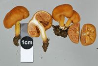
Owner: J.A. Cooper | 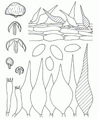
Caption: Fig. 11. Macrocystidia reducta Horak & Capellano (type) : carpophores, spores,
basidia, cheilo- and pleurocystidia, cuticle | 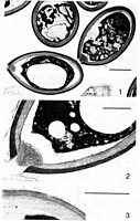
Caption: Macrocystidia reducta HORAK & CAPELLANO (type) : EM-micrographs (taken by A.
CAPELLANO) showing cross-sections of the spores and spore membrane. Scale: 3 µm in
fig. 1; 1 µm in fig. 2 and 3 | 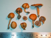
Caption: fruitbody
Owner: J.A. Cooper | 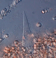
Owner: J.A. Cooper | 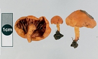
Owner: J.A. Cooper | 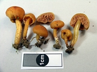
Owner: J.A. Cooper | 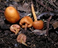
Owner: J.A. Cooper | 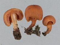
Owner: J.A. Cooper | 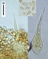
Caption: cystidia
Owner: J.A. Cooper | 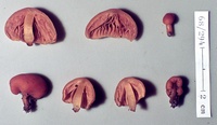
Caption: ZT68-294 , Holotype
Owner: E. Horak: © Creative Commons Attribution-Noncommercial 3.0 New Zealand | 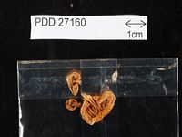
Caption: Dried type specimen
Owner: Herb PDD | |
Article: Horak, E. (1980). New and remarkable Hymenomycetes from tropical forests in Indonesia (Java) and Australasia. Sydowia 33: 39-63.
Description: Pileus -30 mm, ovoid to subglobose when young, inrolled to incurved margin enclosing stipe near
base, becoming pulvinate in aged specimens, never expanded or up-rolled margin; ochre-brown to
orange-brown; dry, smooth to minutely velutinous, margin not striate, not hygrophanous, veil
remnants absent. Lamellae adnexed to adnate, crowded, up to 4 mm wide, straight and not
anastomosing ; pale pink-brown or pale red-brown, edge concolorous, fimbriate. Stipe -15X-4
mm, cylindric, rather short and stout, central; concolorous with pileus; dry, minutely velutinous
all over, becoming fistulose, single in groups. Context pale orange, whitish towards the
centre of pileus and stipe. Odour and taste unpleasant, like rotten fish, rancid. Chemical
reactions on pileus : KOH- negative. Spore print not observed. Spores 7.5-10 X 4.5-5
µm, ovoid to ellipsoid, pale brown, smooth, inamyloid; membrane composed of 4 distinct
layers (pl. 1, fig. 1, 2, 3) viz. rather thick endosporium followed by the strongly developed
sclerosporium with apparent leptotunica, covered here and there by remnants of a loose perisporium
(EM data kindly submitted by A. CAPELLANO, Lyon). Basidia 28-35x6-8 µm, 4-spored. Cheilo-,
pleuro- and caulocystidia 40-90 X 15-30 µm, broadly fusoid with acute apex (awl-shaped),
membrane thin-walled, pale yellow plasmatic pigment present. Cuticle a cutis of cylindric to oval
cells (5-20 µm diam.), with numerous dermato-cystidia morphologically similar to cheilocystidia,
grey-yellow (KOH) pigment dissolved in cell-sap. Clamp connections present.
Habitat: Habitat. - On soil in forests (dominated by Podocarpus dacrydioides, P. ferrugineus, P. spicatus,
Fuchsia excorticata, Melycitus ramiflorus). - New Zealand.
Notes: Macrocystidia JOSSERAND 1933 (HORAK 1968: 360) is a small genus of agarics which are
chiefly characterized by pink to red-brown spore print, ellipsoid and smooth spores and conspicuous
pointed cystidia occurring almost everywhere on the surface of the carpophores. These typical
features are also found in M. reducta. However, the New Zealand representative is separated from
related species by the sub-secotioid carpophores and the ochre-brown colour of pileus and stipe.
The particular micro-structure of the sporal membrane in Macrocystidia (M. cucumis, M.
occidentalis; CAPELLANO 1976) suggested to examine also the spores of M. reducta.
The EM-micrographs revealed that the spore wall is composed of the following distinct layers perisporium,
leptotunica, sclerosporium and endosporium (pl. l, 1-3). This type of structure is also found in the
remaining species of the genus and therefore M. reducta has to be considered a typical member of
Macrocystidia.
|