|
 Conocybe piloselloides Conocybe piloselloides
BiostatusOccurrence uncertain
Images (click to enlarge)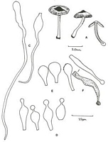
Caption: A, C-F Taylor 1055, A. Habit sketch and section; C. Hairs on stipe; D. Cheilocystidia; E. Pileipellis units; F. Blematogen fragments. |
Article: Watling, R.; Taylor, G.M. (1987). Observations on the Bolbitiaceae: 27. Preliminary account of the Bolbitiaceae of New Zealand. Bibliotheca Mycologica 117: 61 p. + 17 pl.
Description: Description of New Zealand material:
Pileus <35 mm, convex-umbonate to plano-convex but retaining umbo, yellow-brown or tawny brown, drying paler (yellowish), disc rather darker, moist, matt, radially wrinkled about umbo; margin radially striate, splitting. Stipe 35-40 x 3 mm, equal but for abruptly bulbous base, concolorous or paler than pileus, hollow. Gills adnexed, yellow-brown, crowded. Flesh extremely thin in pileus, whitish, distinct from the easily removable stipe, fibrous, very brittle in stipe.
Basidiospores brownish yellow-ochre in mass, 5.5-7(8.5) x 3.5-4.5 µm, elongate-elliptic in face-view, perhaps slightly amygdaliform in side-view, smooth, non-amyloid, slightly thick-walled, ochraceous in water; germ-pore absent. Basidia 4-spored, hyaline, clavate-pedicellate. Hymenophoral trama subparallel. Cheilocystidia lecythiform, 16.5-21.5 x 6-7 µm, capitulum 3.5-4 µm, some with brownish plasmatic pigment in venter in KOH. Pileipellis a palisadoderm of pyriform to ellipsoid-pedicellate cells some with slightly brownish encrustations, 10-15 µm broad, some with plasmatic pigment, hair-like cells absent; pilocystidia detached, either long encrusted hyphae with clamp-connections (17.5-32 x (2.5-) 4.5-6 µm or short chains arising from ± vesiculose cells c. 12 µm broad, usually with swollen capitulum at apex and often containing brown plasmatic pigment. Pileus trama of hyphae with yellow contents in KOH. Stipitipellis of parallel to subparallel cells producing lageniform to cylindric caulocystidia intermixed with long flexuous hair-like cells <60 µm. Clamp-connections present in pileus trama, pileipellis and stipitipellis.
Notes: The pilocystidia are distinctive and possibly represent remnants of the original blematogen as described for C. rickeniana (Singer) P D Orton and C. mesospora (Kuhner ex) Kuhner & Watling (Watling, 1975). Similar structures have not been found previously in C. piloselloides or C. pilosella (Pers.: Fr.) Kuhner and may indicate the New Zealand material represents an independent yet closely related taxon. Strong developments of pilocystidia are found in C. horakii Walling Taylor (q.v.) and in C. dumetorum (Vel.) Svreck
The fungus is easily recognised by the small basidiospores lacking a germ-pore, strongly developed often-coloured pilocystidia and lageniform to cylindric caulocystidia mixed with hair-like cystidia. For these latter cells Walling (1964 et seq.) has erroneously used the term pilocystidia referring to the dermatocystidia of the pileus as pileocystidia. Patrick Barrows (1979) are correct in pointing out that pilocystidia a term introduced by Buller is derived from Greek where pile- refers to head and not hair (Latin). This has led to some confusion; the hair-like cells are so characteristic we believe they should be recognised as a distinct entity.
|