|
 Antrodiella citrea Antrodiella citrea
SynonymsTyromyces semisupinus
Leptoporus coriolus
Polyporus citreus
Tyromyces citreus
Polystictus citreus
BiostatusPresent in region - Indigenous. Non endemic
Images (click to enlarge)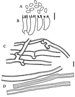
Caption: Fig. 2. Antrodiella citrea. A, basidiospores; B, basidia; C, generative hyphae; D, skeletal
hyphae. A, B, PDD 70909; C, D, PDD 70910. Scale bars = 5 µm. |
Article: Buchanan, P.K.; Ryvarden, L. (2000). New Zealand polypore fungi: six new species and a redetermination. New Zealand Journal of Botany 38(2): 251-263 (http://www.rsnz.org/publish/abstracts.php).
Description: Basidiocarps annual or sometimes reviving for second year, resupinate to effused-reflexed with
lobed pilei, resupinate portion to 9 x 5 cm or sometimes extending along a
branch (e.g., to 20 x 3 cm) but often much smaller, up to 6 mm thick in central part;
reflexed surface cream to bright yellow (86.1.Y - 83.brill.Y), fading in part to yellow- or
orange-brown on drying, scrupose to finely tomentose or glabrous, concentrically zonate;
pore surface bright yellow (83.brill.Y) especially in young material, otherwise cream and
may stain yellow bright yellow colour in part remaining on drying o fading to pale
yellow-brown (73.p.OY), with whit to pale yellow sterile margin to 1 mm wide; pore
angular, with thin dissepiments, 7-9 per mm; tube to 4 mm deep in a layer; context white
though often yellow towards substrate in older specimens, to 1 mm thick. Hyphal system
dimitic; generative hyphae with clamps, hyaline, 2-3.5 µm and thin-walled in trama, 2.5-6 µm diam.
with thickened wall in context, sometimes in context contorted and
frequently branched; skeletal hyphae unbranched, hyaline, thin to thick-walled (to 1.5
µm) and always with distinct lumen, IKI-, with occasional adventitious simple septa, 2-4
µm diam. in trama, 2.5-6 µm diam. in context. Cystidia absent. Basidia clavate, 4-sterigmate,
with a basal clamp, 9-14 x 3.5-4.5 µm. Basidiospores ellipsoid, hyaline, thin-walled, smooth, IKI-,
2.5-3 x 1.2-1.5(-1.8) µm. Causes a white rot, with wood sometimes
staining yellow.
Habitat: SUBSTRATA: On dead hardwoods: Beilschmiedia, Coprosma, Kunzea, and
Leptospermum.
Distribution: New Zealand and Australia.
Notes: NOTES: A. citrea is characterised by a bright yellow, smooth pileus surface, a yellow to
cream pore surface with tiny pores almost invisible macroscopically, and small
basidiospores. Although the species was recorded previously in New Zealand, its
taxonomy has been confused. The description of the type specimen of A. citrea, and
additional material from Australia (Ryvarden 1984) accord with descriptions of New
Zealand and Australian material of Leptoporus coriolus (Reid 1963; Hood 1992) and of
Tyromyces semisupinus sensu G.Cunn. (Cunningham 1965), and is confirmed by our
observation of herbarium material under these names. Collections identified by
Cunningham as T. semisupinus can be readily differentiated from Antrodiella semisupina
(Berk. & M.A.Curtis) Ryvarden (Ryvarden & Gilbertson 1993), and from related species
(Vampola & Pouzar 1996), by their citric yellow pileus surface and the dimitic hyphal
system. A. semisupina sens. str. is not known from New Zealand.
The name Tyromyces citreus (Berk.) G.Cunn. was misapplied by Cunningham (1965) for
New Zealand and Australian collections that differ from A. citrea in having flabelliform
rather than effused-reflexed basidiocarps, and larger pores, basidia, and spores. Further
evaluation of Cunningham's material labelled T. citreus is required (Ryvarden 1984;
Hood 1992).
We consider that the species is correctly placed in Antrodiella Ryvarden & I.Johans.
rather than in Leptoporus Quel. (Reid 1963) since the latter is characterised by a
monomitic hyphal system with simple septate hyphae and a brown rot (Ryvarden 1991).
A. citrea is similar to another species with yellow pores, A. citrinella Niemela &
Ryvarden, but the latter has larger pores, 3-4(-5) per mm, and larger spores, 3-3.5 x 2-2.5
µm (Ryvarden & Gilbertson 1993).
|