|
 Xylaria myosurus Xylaria myosurus
BiostatusOccurrence uncertain
Images (click to enlarge)
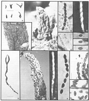
Caption: Fig. 21 A-G Xylaria cf. myosurus A, Stromata. B, C, Stromal surface. D, Ascal ring (Melzer's
reagent). E, Ascospores in an ascus (Melzer's reagent). F, Three ascospores with germ slits
(Melzer's reagent). G, Immature ascospores with a | 
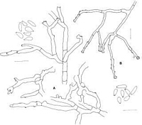
Caption: Fig. 22 Xylaria anamorphs A, X. cf. myosurus (PDD 41977 from culture). B, X.
taxonomic species 1 (PDD 45345 from culture). |
Description: Stromata: Stromata upright (taller than broad) (often branched); stipitate; 7-10 mm tall; 2 mm diam.; perithecia not visible; ostioles finely papillate; stromatal surface wrinkled; dark brown (brown vinaceous), or blackish; KOH-extractable pigments lacking; tissue below perithecia conspicuous, essentially homogeneous, white.
Perithecia: Perithecia more or less globose; 0.1-0.2 mm diam.
Asci: Stipe short with spores filling about two-thirds of ascus; amyloid ring broader than high, or higher than broad.
Ascospores: Ascospores 6-7.5 µm long; 3-4 µm wide; brown; 0-septate; in side view more or less equilateral, or inequilateral, flattened on one side, not curved; in face view elliptic; ends broadly rounded. Germ slit straight, spore-length; on flattened side of spore. Perispore indehiscent in 10% KOH.
Article: Rogers, J.D.; Samuels, G.J. (1987) [1986]. Ascomycetes of New Zealand 8. Xylaria. New Zealand Journal of Botany 24(4): 615-650 (http://www.rsnz.org/publish/abstracts.php).
Description: Stromata gregarious, unbranched or once dichotomously branched, lanceolate to
subcylindrical, 7-10 mm long x 2 mm, widest at base and tapering to an acute tip, elliptical
in section; dark brown to nearly black. Surface rugose but not conspicuously cracked.
Perithecia completely immersed, 100-200 µm diam., perithecial openings appearing as
minute papillae. Internal tissue of stroma white, solid. Stipe lacking or at best ill-defined,
glabrous. Asci 65-95 µm total length x 4-5(-l l) µm, sporiferous part (40-)54-75(-80) µm,
cylindrical; apical ring J+, wedge-shaped, 1 µm high x 1.5 µm wide; ascospores 1-seriate
with overlapping ends. Ascospores (5.0-)6.0-7.5(-8.5) x 3.0-4.0 µm, inequilateral with one
side flat and one side round, elliptic in top view, with inconspicuous cellular appendage
(primary appendage) on one end, transparent brown; slit full length or slightly less, parallel
to long axis of ascospore.
CHARACTERISTICS IN CULTURE: Colonies grown 3 weeks at 15-18°C in diffuse
daylight on OA and CMD c. 1.5 cm diam., felty, white with short aerial hyphae.
Conidiophores formed in poorly developed white hyphal tufts, nondescript, barely
differentiated from vegetative hyphae; Conidia borne along the length on widely spaced,
conspicuous denticles. Conidia (4-)5-7 x 1.5-2.5(-3.0) µm, variable in shape but basically
oblong to clavate, colourless, smooth; each with a protuberant, flat basal abscission scar.
Habitat: HABITAT: On decorticated dicotyledonous trees.
Distribution: DISTRIBUTION: Gisborne.
Notes: Xylaria myosurus has usually been reported from South America. Our concept of
the fungus is based largely on Dennis (1956). The specimens described here are similar to
Dennis' description (1956) except that the ascospores average slightly larger. Type material
[Cayenne, Leprieur (K)] was examined, but ascospores were not sought in the fragile
material.
Martin (1970) cultured ,X. myosurus and his brief description resembles our description,
except that stromata were formed in his cultures. He did not find conidia.
|